
Marine prokaryotes: Difference between revisions
Epipelagic (talk | contribs) →Marine bacteria: from bacteria |
Epipelagic (talk | contribs) |
||
| Line 34: | Line 34: | ||
Microscopic organisms live throughout the [[biosphere]]. The mass of [[prokaryote]] microorganisms — which includes bacteria and archaea, but not the nucleated [[Microorganism#Eukaryotes|eukaryote microorganisms]] — may be as much as 0.8 trillion tons of carbon (of the total biosphere [[Biomass (ecology)|mass]], estimated at between 1 and 4 trillion tons).<ref name="AGCI-2014">{{cite web |author=Staff |title=The Biosphere |url=http://www.agci.org/classroom/biosphere/index.php |date=2014 |work=[[The Given Institute|Aspen Global Change Institute]] |accessdate=10 November 2014}}</ref> |
Microscopic organisms live throughout the [[biosphere]]. The mass of [[prokaryote]] microorganisms — which includes bacteria and archaea, but not the nucleated [[Microorganism#Eukaryotes|eukaryote microorganisms]] — may be as much as 0.8 trillion tons of carbon (of the total biosphere [[Biomass (ecology)|mass]], estimated at between 1 and 4 trillion tons).<ref name="AGCI-2014">{{cite web |author=Staff |title=The Biosphere |url=http://www.agci.org/classroom/biosphere/index.php |date=2014 |work=[[The Given Institute|Aspen Global Change Institute]] |accessdate=10 November 2014}}</ref> |
||
The earliest evidence for life on earth comes from [[Biogenic substance|biogenic]] [[Δ13C|carbon signatures]]<ref name="Rosing 674–676">{{Cite journal|last=Rosing|first=Minik T.|date=1999-01-29|title=13C-Depleted Carbon Microparticles in >3700-Ma Sea-Floor Sedimentary Rocks from West Greenland|journal=Science|language=en|volume=283|issue=5402|pages=674–676|doi=10.1126/science.283.5402.674|issn=0036-8075|pmid=9924024|bibcode=1999Sci...283..674R}}</ref><ref name="Ohtomo 25–28">{{Cite journal|last=Ohtomo|first=Yoko|last2=Kakegawa|first2=Takeshi|last3=Ishida|first3=Akizumi|last4=Nagase|first4=Toshiro|last5=Rosing|first5=Minik T.|date=January 2014|title=Evidence for biogenic graphite in early Archaean Isua metasedimentary rocks|journal=Nature Geoscience|language=En|volume=7|issue=1|pages=25–28|doi=10.1038/ngeo2025|issn=1752-0908|bibcode=2014NatGe...7...25O}}</ref> and [[stromatolite]] fossils<ref>{{Cite journal|last=Nutman|first=Allen P.|last2=Bennett|first2=Vickie C.|last3=Friend|first3=Clark R. L.|last4=Kranendonk|first4=Martin J. Van|last5=Chivas|first5=Allan R.|date=September 2016|title=Rapid emergence of life shown by discovery of 3,700-million-year-old microbial structures|journal=Nature|language=En|volume=537|issue=7621|pages=535–538|doi=10.1038/nature19355|pmid=27580034|issn=1476-4687|bibcode=2016Natur.537..535N|url=http://ro.uow.edu.au/cgi/viewcontent.cgi?article=5181&context=smhpapers}}</ref> discovered in 3.7 billion-year-old [[metasediment]]ary rocks from western [[Greenland]]. In 2015, possible "remains of [[Biotic material|biotic life]]" were found in 4.1 billion-year-old rocks in Western Australia.<ref name="AP-20151019">{{cite news |last=Borenstein |first=Seth |title=Hints of life on what was thought to be desolate early Earth |url=http://apnews.excite.com/article/20151019/us-sci--earliest_life-a400435d0d.html |date=19 October 2015 |work=[[Excite]] |location=Yonkers, NY |publisher=[[Mindspark Interactive Network]] |agency=[[Associated Press]] |url-status=dead |archiveurl=https://web.archive.org/web/20151023200248/http://apnews.excite.com/article/20151019/us-sci--earliest_life-a400435d0d.html |archivedate=23 October 2015 |accessdate=8 October 2018}}</ref><ref name="PNAS-20151014-pdf">{{cite journal |last1=Bell |first1=Elizabeth A. |last2=Boehnike |first2=Patrick |last3=Harrison |first3=T. Mark |last4=Mao |first4=Wendy L. |display-authors=3 |date=19 October 2015 |title=Potentially biogenic carbon preserved in a 4.1 billion-year-old zircon |url=http://www.pnas.org/content/early/2015/10/14/1517557112.full.pdf |journal=Proc. Natl. Acad. Sci. U.S.A. |doi=10.1073/pnas.1517557112 |accessdate=2015-10-20 |pmid=26483481 |pmc=4664351 |volume=112 |issue=47 |pages=14518–21|bibcode=2015PNAS..11214518B }} Early edition, published online before print.</ref> In March 2017, putative evidence of possibly the oldest forms of life on Earth was reported in the form of fossilized [[microorganism]]s discovered in [[hydrothermal vent]] precipitates in the [[Nuvvuagittuq Greenstone Belt|Nuvvuagittuq Belt]] of Quebec, Canada, that may have lived as early as 4.28 billion years ago, not long after the [[Origin of water on Earth#Water in the development of Earth|oceans formed]] 4.4 billion years ago, and not long after the [[Age of the Earth|formation of the Earth]] 4.54 billion years ago.<ref name="NAT-20170301">{{cite journal |author=Dodd, Matthew S. |author2=Papineau, Dominic |author3=Grenne, Tor |author4=slack, John F. |author5=Rittner, Martin |author6=Pirajno, Franco |author7=O'Neil, Jonathan |author8=Little, Crispin T. S. |title=Evidence for early life in Earth's oldest hydrothermal vent precipitates|journal=[[Nature (journal)|Nature]] |volume=543 |issue=7643 |pages=60–64 |date=2 March 2017 |doi=10.1038/nature21377|pmid=28252057 |bibcode=2017Natur.543...60D |url=http://eprints.whiterose.ac.uk/112179/1/ppnature21377_Dodd_for%20Symplectic.pdf }}</ref><ref name="NYT-20170301">{{cite news |last=Zimmer |first=Carl |authorlink=Carl Zimmer |title=Scientists Say Canadian Bacteria Fossils May Be Earth's Oldest |url=https://www.nytimes.com/2017/03/01/science/earths-oldest-bacteria-fossils.html |date=1 March 2017 |work=[[The New York Times]] |accessdate=2 March 2017 }}</ref> |
|||
Microbial mats of coexisting [[bacteria]] and [[archaea]] were the dominant form of life in the early [[Archean]] Epoch and many of the major steps in early evolution are thought to have taken place in this environment.<ref name="NisbetFowler1999ArchaeanMetabolicEvolution">{{cite journal |last1=Nisbet |first1=Euan G. |last2=Fowler |first2=C. M. R. |date=December 7, 1999 |title=Archaean metabolic evolution of microbial mats |journal=[[Proceedings of the Royal Society#Proceedings of the Royal Society B|Proceedings of the Royal Society B]] |volume=266 |issue=1436 |pages=2375–2382 |doi=10.1098/rspb.1999.0934 |pmc=1690475 }}</ref> The evolution of [[photosynthesis]], around 3.5 Ga, eventually led to a buildup of its waste product, [[oxygen]], in the atmosphere, leading to the [[great oxygenation event]], beginning around 2.4 Ga.<ref name="Anbar2007">{{cite journal |last1=Anbar |first1=Ariel D. |author2=Yun Duan |last3=Lyons |first3=Timothy W. |last4=Arnold |first4=Gail L. |last5=Kendall |first5=Brian |last6=Creaser |first6=Robert A. |last7=Kaufman |first7=Alan J. |last8=Gordon |first8=Gwyneth W. |last9=Scott |first9=Clinton |last10=Garvin |first10=Jessica |last11=Buick |first11=Roger |display-authors=3 |date=September 28, 2007 |title=A Whiff of Oxygen Before the Great Oxidation Event? |journal=Science |volume=317 |issue=5846 |pages=1903–1906 |bibcode=2007Sci...317.1903A |doi=10.1126/science.1140325 |pmid=17901330}}</ref> The earliest evidence of [[eukaryote]]s (complex [[Cell (biology)|cell]]s with [[organelle]]s) dates from 1.85 Ga,<ref>{{cite journal |last1=Knoll |first1=Andrew H. |authorlink=Andrew H. Knoll |last2=Javaux |first2=Emmanuelle J. |last3=Hewitt |first3=David |last4=Cohen |first4=Phoebe |title=Eukaryotic organisms in Proterozoic oceans |date=June 29, 2006 |journal=[[Philosophical Transactions of the Royal Society B]] |volume=361 |issue=1470 |pages=1023–1038 |doi=10.1098/rstb.2006.1843 |pmc=1578724 |pmid=16754612 }}</ref><ref name="Fedonkin2003OriginOfMetazoa">{{cite journal |last=Fedonkin |first=Mikhail A. |authorlink=Mikhail Fedonkin |date=March 31, 2003 |title=The origin of the Metazoa in the light of the Proterozoic fossil record |url=http://www.vend.paleo.ru/pub/Fedonkin_2003.pdf |journal=Paleontological Research |volume=7 |issue=1 |pages=9–41 |doi=10.2517/prpsj.7.9 |accessdate=2008-09-02 |url-status=dead |archiveurl=https://web.archive.org/web/20090226122725/http://www.vend.paleo.ru/pub/Fedonkin_2003.pdf |archivedate=February 26, 2009 }}</ref> and while they may have been present earlier, their diversification accelerated when they started using oxygen in their [[metabolism]]. Later, around 1.7 Ga, [[multicellular organism]]s began to appear, with [[Cellular differentiation|differentiated cell]]s performing specialised functions.<ref name="Bonner1999OriginsOfMulticellularity">{{cite journal |last=Bonner |first=John Tyler |authorlink=John Tyler Bonner |year=1998 |title=The origins of multicellularity |journal=[[Integrative Biology]] |volume=1 |issue=1 |pages=27–36 |doi=10.1002/(SICI)1520-6602(1998)1:1<27::AID-INBI4>3.0.CO;2-6 }}</ref> |
|||
==Marine archaea== |
==Marine archaea== |
||
Revision as of 08:24, 8 May 2020
Marine prokaryotes are defined by their habitat as prokaryotes that live in marine environments, that is, in the saltwater of seas or oceans or the brackish water of coastal estuaries. Life forms can be divided into prokaryotes and eukaryotes. Eukaryotes are organisms whose cells have a nucleus enclosed within membranes, whereas prokaryotes have neither a nucleus nor a membrane.[1][2][3] The three-domain system of classifying life adds a third division: the prokaryotes are divided into two domains of life, archaea and bacteria, while the eukaryotes become the third domain.[4]
Background


The word prokaryote comes from the Greek pro meaning "before" and karyon meaning "nut" or "kernel".[5][6] The division between prokaryotes and eukaryotes was firmly established by the microbiologists Roger Stanier and C. B. van Niel in their 1962 paper The concept of a bacterium[7] One reason for this classification was so what was then often called blue-green algae (now called cyanobacteria) would cease to be classified as plants but grouped with bacteria.
In the three-domain system introduced by Carl Woese et al. in 1990[8][9] prokaryotes were further split into into archaea, bacteria domains, while the eukaryote become a domain in their own right. The key difference from earlier classifications is the splitting of archaea from bacteria.
Prokaryotes are asexual, reproducing without fusion of gametes. The first organisms may have been marine prokaryotes.
Prokaryotes can play important roles in nutrient recycling in ecosystems as they act as decomposers. Some prokaryotes are pathogenic, causing disease and even death in plants and animals.[10] Prokaryotes are responsible for significant levels of the photosynthesis that occurs in the ocean, as well as significant cycling of carbon and other nutrients.[11]
Microscopic life undersea is diverse and still poorly understood, such as for the role of viruses in marine ecosystems.[12] Most marine viruses are bacteriophages, which are harmless to plants and animals, but are essential to the regulation of saltwater and freshwater ecosystems.[13] They infect and destroy bacteria in aquatic microbial communities, and are the most important mechanism of recycling carbon in the marine environment. The organic molecules released from the dead bacterial cells stimulate fresh bacterial and algal growth.[14] Viral activity may also contribute to the biological pump, the process whereby carbon is sequestered in the deep ocean.[15]


A stream of airborne microorganisms circles the planet above weather systems but below commercial air lanes.[16] Some peripatetic microorganisms are swept up from terrestrial dust storms, but most originate from marine microorganisms in sea spray. In 2018, scientists reported that hundreds of millions of viruses and tens of millions of bacteria are deposited daily on every square meter around the planet.[17][18]
Microscopic organisms live throughout the biosphere. The mass of prokaryote microorganisms — which includes bacteria and archaea, but not the nucleated eukaryote microorganisms — may be as much as 0.8 trillion tons of carbon (of the total biosphere mass, estimated at between 1 and 4 trillion tons).[19]
The earliest evidence for life on earth comes from biogenic carbon signatures[20][21] and stromatolite fossils[22] discovered in 3.7 billion-year-old metasedimentary rocks from western Greenland. In 2015, possible "remains of biotic life" were found in 4.1 billion-year-old rocks in Western Australia.[23][24] In March 2017, putative evidence of possibly the oldest forms of life on Earth was reported in the form of fossilized microorganisms discovered in hydrothermal vent precipitates in the Nuvvuagittuq Belt of Quebec, Canada, that may have lived as early as 4.28 billion years ago, not long after the oceans formed 4.4 billion years ago, and not long after the formation of the Earth 4.54 billion years ago.[25][26]
Microbial mats of coexisting bacteria and archaea were the dominant form of life in the early Archean Epoch and many of the major steps in early evolution are thought to have taken place in this environment.[27] The evolution of photosynthesis, around 3.5 Ga, eventually led to a buildup of its waste product, oxygen, in the atmosphere, leading to the great oxygenation event, beginning around 2.4 Ga.[28] The earliest evidence of eukaryotes (complex cells with organelles) dates from 1.85 Ga,[29][30] and while they may have been present earlier, their diversification accelerated when they started using oxygen in their metabolism. Later, around 1.7 Ga, multicellular organisms began to appear, with differentiated cells performing specialised functions.[31]
Marine archaea

The archaea (Greek for ancient[33]) constitute a domain and kingdom of single-celled microorganisms. These microbes are prokaryotes, meaning they have no cell nucleus or any other membrane-bound organelles in their cells.
Archaea were initially classified as bacteria, but this classification is outdated.[34] Archaeal cells have unique properties separating them from the other two domains of life, Bacteria and Eukaryota. The Archaea are further divided into multiple recognized phyla. Classification is difficult because the majority have not been isolated in the laboratory and have only been detected by analysis of their nucleic acids in samples from their environment.
Archaea and bacteria are generally similar in size and shape, although a few archaea have very strange shapes, such as the flat and square-shaped cells of Haloquadratum walsbyi.[35] Despite this morphological similarity to bacteria, archaea possess genes and several metabolic pathways that are more closely related to those of eukaryotes, notably the enzymes involved in transcription and translation. Other aspects of archaeal biochemistry are unique, such as their reliance on ether lipids in their cell membranes, such as archaeols. Archaea use more energy sources than eukaryotes: these range from organic compounds, such as sugars, to ammonia, metal ions or even hydrogen gas. Salt-tolerant archaea (the Haloarchaea) use sunlight as an energy source, and other species of archaea fix carbon; however, unlike plants and cyanobacteria, no known species of archaea does both. Archaea reproduce asexually by binary fission, fragmentation, or budding; unlike bacteria and eukaryotes, no known species forms spores.
Archaea are particularly numerous in the oceans, and the archaea in plankton may be one of the most abundant groups of organisms on the planet. Archaea are a major part of Earth's life and may play roles in both the carbon cycle and the nitrogen cycle. Crenarchaeota (eocytes) are a phylum of archaea thought to be very abundant in marine environments and one of the main contributors to the fixation of carbon.[36]
-
 Eocytes may be the most abundant of marine archaea
 -
 Halobacteria, found in water near saturated with salt, are now recognised as archaea.
 -
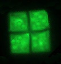 Flat, square-shaped cells of the archaea Haloquadratum walsbyi
 -
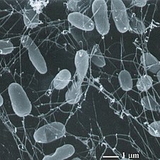
 Methanosarcina barkeri, a marine archaea that produces methane
 -
 Thermophiles, such as Pyrolobus fumarii, survive well over 100 °C


Maine archaea may be classified as follows:[37][38][39][40][41]
- Marine Group I (MG-I or MGI): marine Thaumarchaeota with subgroups Ia (aka I.a) up to Id
- Marine Group II (MG-II): marine Euryarchaeota, order Poseidoniales[42] with subgroups IIa up to IId (IIa resembling Poseidoniaceae, IIb resembling Thalassarchaceae)
Viruses parasiting MGII are classified as magroviruses - Marine Group III (MG-III): also marine Euryarchaeota, Marine Benthic Group D[43]
- Marine Group IV (MG-IV): also marine Euryarchaeota[44]
Marine bacteria


Bacteria constitute a large domain of prokaryotic microorganisms. Typically a few micrometres in length, bacteria have a number of shapes, ranging from spheres to rods and spirals. Bacteria were among the first life forms to appear on Earth, and are present in most of its habitats. Bacteria inhabit soil, water, acidic hot springs, radioactive waste,[46] and the deep portions of Earth's crust. Bacteria also live in symbiotic and parasitic relationships with plants and animals.
Once regarded as plants constituting the class Schizomycetes, bacteria are now classified as prokaryotes. Unlike cells of animals and other eukaryotes, bacterial cells do not contain a nucleus and rarely harbour membrane-bound organelles. Although the term bacteria traditionally included all prokaryotes, the scientific classification changed after the discovery in the 1990s that prokaryotes consist of two very different groups of organisms that evolved from an ancient common ancestor. These evolutionary domains are called Bacteria and Archaea.[47]
The ancestors of modern bacteria were unicellular microorganisms that were the first forms of life to appear on Earth, about 4 billion years ago. For about 3 billion years, most organisms were microscopic, and bacteria and archaea were the dominant forms of life.[48][49] Although bacterial fossils exist, such as stromatolites, their lack of distinctive morphology prevents them from being used to examine the history of bacterial evolution, or to date the time of origin of a particular bacterial species. However, gene sequences can be used to reconstruct the bacterial phylogeny, and these studies indicate that bacteria diverged first from the archaeal/eukaryotic lineage.[50] Bacteria were also involved in the second great evolutionary divergence, that of the archaea and eukaryotes. Here, eukaryotes resulted from the entering of ancient bacteria into endosymbiotic associations with the ancestors of eukaryotic cells, which were themselves possibly related to the Archaea.[51][52] This involved the engulfment by proto-eukaryotic cells of alphaproteobacterial symbionts to form either mitochondria or hydrogenosomes, which are still found in all known Eukarya. Later on, some eukaryotes that already contained mitochondria also engulfed cyanobacterial-like organisms. This led to the formation of chloroplasts in algae and plants. There are also some algae that originated from even later endosymbiotic events. Here, eukaryotes engulfed a eukaryotic algae that developed into a "second-generation" plastid.[53][54] This is known as secondary endosymbiosis.
Bacteria grow to a fixed size and then reproduce through binary fission, a form of asexual reproduction.[55] Under optimal conditions, bacteria can grow and divide extremely rapidly, and bacterial populations can double as quickly as every 9.8 minutes.[56]
Pelagibacter ubique and its relatives may be the most abundant microorganisms in the ocean, and it has been claimed that they are possibly the most abundant bacteria in the world. They make up about 25% of all microbial plankton cells, and in the summer they may account for approximately half the cells present in temperate ocean surface water. The total abundance of P. ubique and relatives is estimated to be about 2 × 1028 microbes.[57] However, it was reported in Nature in February 2013 that the bacteriophage HTVC010P, which attacks P. ubique, has been discovered and is probably the most common organism on the planet.[58][59]
Roseobacter is also one of the most abundant and versatile microorganisms in the ocean. They are diversified across different types of marine habitats, from coastal to open oceans and from sea ice to sea floor, and make up about 25% of coastal marine bacteria. Members of the Roseobacter genus play important roles in marine biogeochemical cycles and climate change, processing a significant portion of the total carbon in the marine environment. They form symbiotic relationships which allow them to degrade aromatic compounds and uptake trace metals. They are widely used in aquaculture and quorum sensing. During algal blooms, 20-30% of the prokaryotic community are Roseobacter.[60][61]
The largest known bacterium, the marine Thiomargarita namibiensis, can be visible to the naked eye and sometimes attains 0.75 mm (750 μm).[62][63]
-
 The marine Thiomargarita namibiensis, largest known bacterium
 -
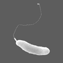 The bacterium Marinomonas arctica grows inside Arctic sea ice at subzero temperatures


- Aerobic anoxygenic phototrophic bacteria (AAPBs) are widely distributed marine plankton that may constitute over 10% of the open ocean microbial community. Marine AAPBs are classified in two marine (Erythrobacter and Roseobacter) genera. They can be particularly abundant in oligotrophic conditions where they were found to be 24% of the community.[64] These are heterotrophic organisms that use light to produce energy, but are unable to utilise carbon dioxide as their primary carbon source. Most are obligately aerobic, meaning they require oxygen to grow. Current data suggests that marine bacteria have generation times of several days, whereas new evidence exists that shows AAPB to have a much shorter generation time.[65] Coastal/shelf waters often have greater amounts of AAPBs, some as high as 13.51% AAPB%. Phytoplankton also affect AAPB%, but little research has been performed in this area.[66] They can also be abundant in various oligotrophic conditions, including the most oligotrophic regime of the world ocean.[67] They are globally distributed in the euphotic zone and represent a hitherto unrecognized component of the marine microbial community that appears to be critical to the cycling of both organic and inorganic carbon in the ocean.[68]
- Zetaproteobacteria: are iron-oxidizing neutrophilic chemolithoautotrophs, distributed worldwide in estuaries and marine habitats.
- Hydrogen oxidizing bacteria are facultative autotrophs that can be divided into aerobes and anaerobes. The former use hydrogen as an electron donor and oxygen as an acceptor while the latter use sulphate or nitrogen dioxide as electron acceptors.[69]
- Phototrophy
- Phototrophy is a noticeable and significant marker that should always play a primary role in bacterial classification.[70]
Cyanobacteria

.jpg)

| External videos | |
|---|---|
Cyanobacteria were the first organisms to evolve an ability to turn sunlight into chemical energy. They form a phylum (division) of bacteria which range from unicellular to filamentous and include colonial species. They are found almost everywhere on earth: in damp soil, in both freshwater and marine environments, and even on Antarctic rocks.[72] In particular, some species occur as drifting cells floating in the ocean, and as such were amongst the first of the phytoplankton.
The first primary producers that used photosynthesis were oceanic cyanobacteria about 2.3 billion years ago.[73][74] The release of molecular oxygen by cyanobacteria as a by-product of photosynthesis induced global changes in the Earth's environment. Because oxygen was toxic to most life on Earth at the time, this led to the near-extinction of oxygen-intolerant organisms, a dramatic change which redirected the evolution of the major animal and plant species.[75]
The tiny (0.6 µm) marine cyanobacterium Prochlorococcus, discovered in 1986, forms today an important part of the base of the ocean food chain and accounts for much of the photosynthesis of the open ocean[76] and an estimated 20% of the oxygen in the Earth's atmosphere.[77] It is possibly the most plentiful genus on Earth: a single millilitre of surface seawater may contain 100,000 cells or more.[78]
Originally, biologists classified cyanobacteria as an algae, and referred to it as "blue-green algae". The more recent view is that cyanobacteria is a bacteria, and hence is not even in the same Kingdom as algae. Most authorities exclude all prokaryotes, and hence cyanobacteria from the definition of algae.[79][80]
-
-
![The chloroplasts of glaucophytes have a peptidoglycan layer, evidence suggesting their endosymbiotic origin from cyanobacteria.[81]](//upload.wikimedia.org/wikipedia/commons/thumb/c/c9/Glaucocystis_sp.jpg/280px-Glaucocystis_sp.jpg) The chloroplasts of glaucophytes have a peptidoglycan layer, evidence suggesting their endosymbiotic origin from cyanobacteria.[81]
The chloroplasts of glaucophytes have a peptidoglycan layer, evidence suggesting their endosymbiotic origin from cyanobacteria.[81]

![The chloroplasts of glaucophytes have a peptidoglycan layer, evidence suggesting their endosymbiotic origin from cyanobacteria.[81]](/wiki/File:Glaucocystis_sp.jpg)
Symbiosis
Some marine organisms have a symbiosis with bacteria or archaea. Pompeii worms live at great depths by hydrothermal vents at temperatures up to 80°C. They have "hairy" backs; these "hairs" are actually colonies of bacteria such as Nautilia profundicola, which are thought to afford the worm some degree of insulation. Glands on the worm's back secrete a mucus on which the bacteria feed, a form of symbiosis.
-
 The "hairy" backs of Pompeii worms are actually colonies of symbiotic bacteria
 -
![Hesiocaeca methanicola lives at great depths on methane ice and appear to survive in symbiosis with bacteria which metabolize the clathrate.[82]](//upload.wikimedia.org/wikipedia/commons/thumb/b/bb/Hesiocaeca_methanicola_noaa.jpg/361px-Hesiocaeca_methanicola_noaa.jpg) Hesiocaeca methanicola lives at great depths on methane ice and appear to survive in symbiosis with bacteria which metabolize the clathrate.[82]
Hesiocaeca methanicola lives at great depths on methane ice and appear to survive in symbiosis with bacteria which metabolize the clathrate.[82]

![Hesiocaeca methanicola lives at great depths on methane ice and appear to survive in symbiosis with bacteria which metabolize the clathrate.[82]](/wiki/File:Hesiocaeca_methanicola_noaa.jpg)
Microbial rhodopsin

(2) it changes its configuration so a proton is expelled from the cell
(3) the chemical potential causes the proton to flow back to the cell
(4) thus generating energy
(5) in the form of adenosine triphosphate.[83]
Phototrophic metabolism relies on one of three energy-converting pigments: chlorophyll, bacteriochlorophyll, and retinal. Retinal is the chromophore found in rhodopsins. The significance of chlorophyll in converting light energy has been written about for decades, but phototrophy based on retinal pigments is just beginning to be studied.[84]
.jpg)
In 2000 a team of microbiologists led by Edward DeLong made a crucial discovery in the understanding of the marine carbon and energy cycles. They discovered a gene in several species of bacteria[86][87] responsible for production of the protein rhodopsin, previously unheard of in bacteria. These proteins found in the cell membranes are capable of converting light energy to biochemical energy due to a change in configuration of the rhodopsin molecule as sunlight strikes it, causing the pumping of a proton from inside out and a subsequent inflow that generates the energy.[88] The archaeal-like rhodopsins have subsequently been found among different taxa, protists as well as in bacteria and archaea, though they are rare in complex multicellular organisms.[89][90][91]
Research in 2019 shows these "sun-snatching bacteria" are more widespread than previously thought and could change how oceans are affected by global warming. "The findings break from the traditional interpretation of marine ecology found in textbooks, which states that nearly all sunlight in the ocean is captured by chlorophyll in algae. Instead, rhodopsin-equipped bacteria function like hybrid cars, powered by organic matter when available — as most bacteria are — and by sunlight when nutrients are scarce."[92][84]
There is an astrobiological conjecture called the Purple Earth hypothesis which surmises that original life forms on Earth were retinal-based rather than chlorophyll-based, which would have made the Earth appear purple instead of green.[93][94]
Role in chemical cycling
Archaea recycle elements such as carbon, nitrogen, and sulfur through their various habitats.[95] Archaea carry out many steps in the nitrogen cycle. This includes both reactions that remove nitrogen from ecosystems (such as nitrate-based respiration and denitrification) as well as processes that introduce nitrogen (such as nitrate assimilation and nitrogen fixation).[96][97]
Researchers recently discovered archaeal involvement in ammonia oxidation reactions. These reactions are particularly important in the oceans.[98][99] In the sulfur cycle, archaea that grow by oxidizing sulfur compounds release this element from rocks, making it available to other organisms, but the archaea that do this, such as Sulfolobus, produce sulfuric acid as a waste product, and the growth of these organisms in abandoned mines can contribute to acid mine drainage and other environmental damage.[100] In the carbon cycle, methanogen archaea remove hydrogen and play an important role in the decay of organic matter by the populations of microorganisms that act as decomposers in anaerobic ecosystems, such as sediments and marshes.[101]
References
- ^ Youngson, Robert M. (2006). Collins Dictionary of Human Biology. Glasgow: HarperCollins. ISBN 978-0-00-722134-9.
{{cite book}}: Unknown parameter|name-list-format=ignored (|name-list-style=suggested) (help) - ^ Nelson, David L.; Cox, Michael M. (2005). Lehninger Principles of Biochemistry (4th ed.). New York: W.H. Freeman. ISBN 978-0-7167-4339-2.
{{cite book}}: Unknown parameter|name-list-format=ignored (|name-list-style=suggested) (help) - ^ Martin EA, ed. (1983). Macmillan Dictionary of Life Sciences (2nd ed.). London: Macmillan Press. ISBN 978-0-333-34867-3.
- ^ Coté, Gary; De Tullio, Mario (2010). "Beyond Prokaryotes and Eukaryotes: Planctomycetes and Cell Organization". Nature.
{{cite journal}}: Unknown parameter|name-list-format=ignored (|name-list-style=suggested) (help) - ^ Campbell, N. "Biology:Concepts & Connections". Pearson Education. San Francisco: 2003.
- ^ "prokaryote". Online Etymology Dictionary.
- ^ Stanier RY, Van Niel CB (1962). "The concept of a bacterium". Archiv für Mikrobiologie. 42: 17–35. doi:10.1007/BF00425185. PMID 13916221.
- ^ Woese CR, Fox GE (November 1977). "Phylogenetic structure of the prokaryotic domain: the primary kingdoms". Proceedings of the National Academy of Sciences of the United States of America. 74 (11): 5088–90. Bibcode:1977PNAS...74.5088W. doi:10.1073/pnas.74.11.5088. PMC 432104. PMID 270744.
- ^ Woese CR, Kandler O, Wheelis ML (June 1990). "Towards a natural system of organisms: proposal for the domains Archaea, Bacteria, and Eucarya". Proceedings of the National Academy of Sciences of the United States of America. 87 (12): 4576–9. Bibcode:1990PNAS...87.4576W. doi:10.1073/pnas.87.12.4576. PMC 54159. PMID 2112744.
- ^ 2002 WHO mortality data Accessed 20 January 2007
- ^ "Functions of global ocean microbiome key to understanding environmental changes". www.sciencedaily.com. University of Georgia. 10 December 2015. Retrieved 11 December 2015.
- ^ Suttle, C.A. (2005). "Viruses in the Sea". Nature. 437 (9): 356–361. Bibcode:2005Natur.437..356S. doi:10.1038/nature04160. PMID 16163346.
- ^ Shors p. 5
- ^ Shors p. 593
- ^ Suttle CA. Marine viruses—major players in the global ecosystem. Nature Reviews Microbiology. 2007;5(10):801–12. doi:10.1038/nrmicro1750. PMID 17853907.
- ^ Living Bacteria Are Riding Earth’s Air Currents Smithsonian Magazine, 11 January 2016.
- ^ Robbins, Jim (13 April 2018). "Trillions Upon Trillions of Viruses Fall From the Sky Each Day". The New York Times. Retrieved 14 April 2018.
- ^ Reche, Isabel; D’Orta, Gaetano; Mladenov, Natalie; Winget, Danielle M; Suttle, Curtis A (29 January 2018). "Deposition rates of viruses and bacteria above the atmospheric boundary layer". ISME Journal. 12 (4): 1154–1162. doi:10.1038/s41396-017-0042-4. PMC 5864199. PMID 29379178.
- ^ Staff (2014). "The Biosphere". Aspen Global Change Institute. Retrieved 10 November 2014.
- ^ Rosing, Minik T. (1999-01-29). "13C-Depleted Carbon Microparticles in >3700-Ma Sea-Floor Sedimentary Rocks from West Greenland". Science. 283 (5402): 674–676. Bibcode:1999Sci...283..674R. doi:10.1126/science.283.5402.674. ISSN 0036-8075. PMID 9924024.
- ^ Ohtomo, Yoko; Kakegawa, Takeshi; Ishida, Akizumi; Nagase, Toshiro; Rosing, Minik T. (January 2014). "Evidence for biogenic graphite in early Archaean Isua metasedimentary rocks". Nature Geoscience. 7 (1): 25–28. Bibcode:2014NatGe...7...25O. doi:10.1038/ngeo2025. ISSN 1752-0908.
- ^ Nutman, Allen P.; Bennett, Vickie C.; Friend, Clark R. L.; Kranendonk, Martin J. Van; Chivas, Allan R. (September 2016). "Rapid emergence of life shown by discovery of 3,700-million-year-old microbial structures". Nature. 537 (7621): 535–538. Bibcode:2016Natur.537..535N. doi:10.1038/nature19355. ISSN 1476-4687. PMID 27580034.
- ^ Borenstein, Seth (19 October 2015). "Hints of life on what was thought to be desolate early Earth". Excite. Yonkers, NY: Mindspark Interactive Network. Associated Press. Archived from the original on 23 October 2015. Retrieved 8 October 2018.
- ^ Bell, Elizabeth A.; Boehnike, Patrick; Harrison, T. Mark; et al. (19 October 2015). "Potentially biogenic carbon preserved in a 4.1 billion-year-old zircon" (PDF). Proc. Natl. Acad. Sci. U.S.A. 112 (47): 14518–21. Bibcode:2015PNAS..11214518B. doi:10.1073/pnas.1517557112. PMC 4664351. PMID 26483481. Retrieved 2015-10-20. Early edition, published online before print.
- ^ Dodd, Matthew S.; Papineau, Dominic; Grenne, Tor; slack, John F.; Rittner, Martin; Pirajno, Franco; O'Neil, Jonathan; Little, Crispin T. S. (2 March 2017). "Evidence for early life in Earth's oldest hydrothermal vent precipitates" (PDF). Nature. 543 (7643): 60–64. Bibcode:2017Natur.543...60D. doi:10.1038/nature21377. PMID 28252057.
- ^ Zimmer, Carl (1 March 2017). "Scientists Say Canadian Bacteria Fossils May Be Earth's Oldest". The New York Times. Retrieved 2 March 2017.
- ^ Nisbet, Euan G.; Fowler, C. M. R. (December 7, 1999). "Archaean metabolic evolution of microbial mats". Proceedings of the Royal Society B. 266 (1436): 2375–2382. doi:10.1098/rspb.1999.0934. PMC 1690475.
- ^ Anbar, Ariel D.; Yun Duan; Lyons, Timothy W.; et al. (September 28, 2007). "A Whiff of Oxygen Before the Great Oxidation Event?". Science. 317 (5846): 1903–1906. Bibcode:2007Sci...317.1903A. doi:10.1126/science.1140325. PMID 17901330.
- ^ Knoll, Andrew H.; Javaux, Emmanuelle J.; Hewitt, David; Cohen, Phoebe (June 29, 2006). "Eukaryotic organisms in Proterozoic oceans". Philosophical Transactions of the Royal Society B. 361 (1470): 1023–1038. doi:10.1098/rstb.2006.1843. PMC 1578724. PMID 16754612.
- ^ Fedonkin, Mikhail A. (March 31, 2003). "The origin of the Metazoa in the light of the Proterozoic fossil record" (PDF). Paleontological Research. 7 (1): 9–41. doi:10.2517/prpsj.7.9. Archived from the original (PDF) on February 26, 2009. Retrieved 2008-09-02.
- ^ Bonner, John Tyler (1998). "The origins of multicellularity". Integrative Biology. 1 (1): 27–36. doi:10.1002/(SICI)1520-6602(1998)1:1<27::AID-INBI4>3.0.CO;2-6.
- ^ Bang C, Schmitz RA (2015). "Archaea associated with human surfaces: not to be underestimated". FEMS Microbiology Reviews. 39 (5): 631–48. doi:10.1093/femsre/fuv010. PMID 25907112.
- ^ Archaea Online Etymology Dictionary. Retrieved 17 August 2016.
- ^ Pace NR (May 2006). "Time for a change". Nature. 441 (7091): 289. Bibcode:2006Natur.441..289P. doi:10.1038/441289a. PMID 16710401.
- ^ Stoeckenius W (1 October 1981). "Walsby's square bacterium: fine structure of an orthogonal procaryote". Journal of Bacteriology. 148 (1): 352–60. doi:10.1128/JB.148.1.352-360.1981. PMC 216199. PMID 7287626.
- ^ Madigan M and Martinko J (Eds.) (2005). Brock Biology of Microorganisms (11th ed.). Prentice Hall. ISBN 978-0-13-144329-7.
- ^ Luis H. Orellana, T. Ben Francis, Karen Krüger, Hanno Teeling, Marie-Caroline Müller, Bernhard M. Fuchs, Konstantinos T. Konstantinidis, Rudolf I. Amann: Niche differentiation among annually recurrent coastal Marine Group II Euryarchaeota. In: Nature ISME Journal 13, pp. 3014-3036, 26 August 2019, doi:10.1038/s41396-019-0491-z
- ^ Yosuke Nishimura, Hiroyasu Watai, Takashi Honda, Tomoko Mihara, Kimiho Omae, Simon Roux, Romain Blanc-Mathieu, Keigo Yamamoto, Pascal Hingamp, Yoshihiko Sako, Matthew B. Sullivan, Susumu Goto, Hiroyuki Ogata, Takashi Yoshidacorresponding: Environmental Viral Genomes Shed New Light on Virus-Host Interactions in the Ocean. In: mSphere 2(2), March–April 2017, e00359-16, doi:10.1128/mSphere.00359-16, PMC 5332604, PMID 28261669, esp. Fig. 4
- ^ Alon Philosof, Natalya Yutin, José Flores-Uribe, Itai Sharon, Eugene V. Koonin, Oded Béjà: Novel Abundant Oceanic Viruses of Uncultured Marine Group II Euryarchaeota. In: Curr Biol. 27(9), 8 May 2017, pp. 1362–1368, doi:10.1016/j.cub.2017.03.052, PMC 5434244, PMID 28457865
- ^ Xiaomin Xia, Wang Guo, Hongbin Liu: Basin Scale Variation on the Composition and Diversity of Archaea in the Pacific Ocean. In: Front. Microbiol., 23 October 2017, doi:10.3389/fmicb.2017.02057
- ^ Ana-Belen Martin-Cuadrado et al.: A new class of marine Euryarchaeota group II from the mediterranean deep chlorophyll maximum. In: Nature ISME Journal volume 9, pp. 1619–1634 (2015), 23 December 2014, doi:10.1038/ismej.2014.249
- ^ NCBI: Candidatus Poseidoniales (order)
- ^ NCBI: Marine Group III
- ^ NCBI: Marine Group IV
- ^ Durham, B.P., Grote, J., Whittaker, K.A., Bender, S.J., Luo, H., Grim, S.L., Brown, J.M., Casey, J.R., Dron, A., Florez-Leiva, L. and Krupke, A. (2014) "Draft genome sequence of marine alphaproteobacterial strain HIMB11, the first cultivated representative of a unique lineage within the Roseobacter clade possessing an unusually small genome". Standards in genomic sciences, 9(3): 632. doi:10.4056/sigs.4998989.
- ^ Fredrickson JK, Zachara JM, Balkwill DL, Kennedy D, Li SM, Kostandarithes HM, Daly MJ, Romine MF, Brockman FJ (2004). "Geomicrobiology of high-level nuclear waste-contaminated vadose sediments at the Hanford site, Washington state". Applied and Environmental Microbiology. 70 (7): 4230–41. doi:10.1128/AEM.70.7.4230-4241.2004. PMC 444790. PMID 15240306.
- ^ Woese CR, Kandler O, Wheelis ML (1990). "Towards a natural system of organisms: proposal for the domains Archaea, Bacteria, and Eucarya". Proceedings of the National Academy of Sciences of the United States of America. 87 (12): 4576–9. Bibcode:1990PNAS...87.4576W. doi:10.1073/pnas.87.12.4576. PMC 54159. PMID 2112744.
- ^ Schopf JW (1994). "Disparate rates, differing fates: tempo and mode of evolution changed from the Precambrian to the Phanerozoic". Proceedings of the National Academy of Sciences of the United States of America. 91 (15): 6735–42. Bibcode:1994PNAS...91.6735S. doi:10.1073/pnas.91.15.6735. PMC 44277. PMID 8041691.
- ^ DeLong EF, Pace NR (2001). "Environmental diversity of bacteria and archaea". Systematic Biology. 50 (4): 470–8. CiteSeerX 10.1.1.321.8828. doi:10.1080/106351501750435040. PMID 12116647.
- ^ Brown JR, Doolittle WF (1997). "Archaea and the prokaryote-to-eukaryote transition". Microbiology and Molecular Biology Reviews. 61 (4): 456–502. doi:10.1128/.61.4.456-502.1997. PMC 232621. PMID 9409149.
- ^ Dyall, Sabrina D.; Brown, Mark T.; Johnson, Patricia J. (9 April 2004). "Ancient Invasions: From Endosymbionts to Organelles". Science. 304 (5668): 253–257. Bibcode:2004Sci...304..253D. doi:10.1126/science.1094884. ISSN 0036-8075. PMID 15073369.
- ^ Poole AM, Penny D (2007). "Evaluating hypotheses for the origin of eukaryotes". BioEssays. 29 (1): 74–84. doi:10.1002/bies.20516. PMID 17187354.
- ^ Lang BF, Gray MW, Burger G (1999). "Mitochondrial genome evolution and the origin of eukaryotes". Annual Review of Genetics. 33: 351–97. doi:10.1146/annurev.genet.33.1.351. PMID 10690412.
- ^ McFadden GI (1999). "Endosymbiosis and evolution of the plant cell". Current Opinion in Plant Biology. 2 (6): 513–9. doi:10.1016/S1369-5266(99)00025-4. PMID 10607659.
- ^ Koch AL (2002). "Control of the bacterial cell cycle by cytoplasmic growth". Critical Reviews in Microbiology. 28 (1): 61–77. doi:10.1080/1040-840291046696. PMID 12003041.
- ^ Eagon RG (April 1962). "Pseudomonas natriegens, a marine bacterium with a generation time of less than 10 minutes". Journal of Bacteriology. 83 (4): 736–37. doi:10.1128/jb.83.4.736-737.1962. PMC 279347. PMID 13888946.
- ^ "Candidatus Pelagibacter Ubique." European Bioinformatics Institute. European Bioinformatics Institute, 2011. Web. 08 Jan. 2012. http://www.ebi.ac.uk/2can/genomes/bacteria/Candidatus_Pelagibacter_ubique.html Archived December 1, 2008, at the Wayback Machine
- ^ "Flea market: A newly discovered virus may be the most abundant organism on the planet". The Economist. 16 February 2013. Retrieved 16 February 2013.
- ^ Zhao, Y.; Temperton, B.; Thrash, J. C.; Schwalbach, M. S.; Vergin, K. L.; Landry, Z. C.; Ellisman, M.; Deerinck, T.; Sullivan, M. B.; Giovannoni, S. J. (2013). "Abundant SAR11 viruses in the ocean". Nature. 494 (7437): 357–360. Bibcode:2013Natur.494..357Z. doi:10.1038/nature11921. PMID 23407494.
- ^ Bentzon-Tilia, Mikkel; Gram, Lone (2017). Bioprospecting. Topics in Biodiversity and Conservation. Springer, Cham. pp. 137–166. doi:10.1007/978-3-319-47935-4_7. ISBN 978-3-319-47933-0.
{{cite book}}: Unknown parameter|name-list-format=ignored (|name-list-style=suggested) (help) - ^ NCBI Taxonomy Browser: Roseobacter National Center for Biotechnology Information. Accessed: 8 May 2020.
- ^ "The largest Bacterium: Scientist discovers new bacterial life form off the African coast", Max Planck Institute for Marine Microbiology, 8 April 1999, archived from the original on 20 January 2010
- ^ List of Prokaryotic names with Standing in Nomenclature - Genus Thiomargarita
- ^ Lami, R.; Cottrell, M. T.; Ras, J.; Ulloa, O.; Obernosterer, I.; Claustre, H.; Kirchman, D. L.; Lebaron, P. (2007). "High Abundances of Aerobic Anoxygenic Photosynthetic Bacteria in the South Pacific Ocean". Applied and Environmental Microbiology. 73 (13): 4198–205. doi:10.1128/AEM.02652-06. PMC 1932784. PMID 17496136.
- ^ Life science weekly. (2012). Bacteria; Reports from Spanish National Research Council (CSIC) Describe Recent Advances in Bacteria. ISSN 1552-2466. P.4582.
- ^ Jiao, Nianzhi, Zhang, Yao, Zeng, Yonghui, Hong, Ning, Liu, Rulong, Chen, Feng, & Wang, Pinxian (2007). Distinct distribution pattern of abundance and diversity of aerobic anoxygenic phototrophic bacteria in the global ocean. Environmental Microbiology: 9(12), pp.3091-3099
- ^ Rapheal Lami, Matthew T. Cottell, Josephine Ras, Osvaldo Ulloa, Ingrid Obernosterer, Herve Claistre, David L. Kirchman, Philippe Lebaron (2007). "High Abundances of Aerobic Anoxygenic Photosynthetic Bacteria in the South Pacific Ocean". Applied and Environmental Microbiology 73 (13), 4198-4205. doi:10.1128/AEM.02652-06
- ^ Zbigniew S. Kolber, F. Gerald, Plumley, Andrew S. Lang, J. Thomas beatty, Robert E. Blankenship, Cindy L. Vandover, Costantino Vetriani, Michal Koblizek, Christopher Rathgeber, Paul G. Falkowsik (2001). "Contribution of Aerobic Photoheterotrophic Bacteria to the Carbon Cycle in the Ocean". Science 29: 2492-2495.
- ^ Aragno, Michel; Schlegel, Hans G. (1981). "The Hydrogen-Oxidizing Bacteria". In Starr, Mortimer P.; Stolp, Heinz; Trüper, Hans G.; Balows, Albert; Schlegel, Hans G. (eds.). The Prokaryotes. Berlin, Heidelberg: Springer. pp. 865–893. doi:10.1007/978-3-662-13187-9_70. ISBN 978-3-662-13187-9.
{{cite book}}: Unknown parameter|name-list-format=ignored (|name-list-style=suggested) (help) - ^ Yurkov, Vladimir V., & Beatty, Thomas J. (1998). Aerobic Anoxygenic Phototrophic Bacteria. Microbiol Mol Biol Rev. 1998 September; 62(3): 695–724.
- ^ Changes in oxygen concentrations in our ocean can disrupt fundamental biological cycles Phys.org, 25 November 2019.
- ^ Walsh, Patrick J.; Smith, Sharon; Fleming, Lora; Solo-Gabriele, Helena; Gerwick, William H., eds. (2 September 2011). "Cyanobacteria and cyanobacterial toxins". Oceans and Human Health: Risks and Remedies from the Seas. Academic Press. pp. 271–296. ISBN 978-0-08-087782-2.
{{cite book}}: Unknown parameter|name-list-format=ignored (|name-list-style=suggested) (help) - ^ "The Rise of Oxygen - Astrobiology Magazine". Astrobiology Magazine. Retrieved 2016-04-06.
- ^ Flannery, D. T.; R.M. Walter (2012). "Archean tufted microbial mats and the Great Oxidation Event: new insights into an ancient problem". Australian Journal of Earth Sciences. 59 (1): 1–11. Bibcode:2012AuJES..59....1F. doi:10.1080/08120099.2011.607849.
- ^ Rothschild, Lynn (September 2003). "Understand the evolutionary mechanisms and environmental limits of life". NASA. Archived from the original on 11 March 2012. Retrieved 13 July 2009.
- ^ Nadis S (December 2003). "The cells that rule the seas" (PDF). Scientific American. 289 (6): 52–3. Bibcode:2003SciAm.289f..52N. doi:10.1038/scientificamerican1203-52. PMID 14631732. Archived from the original (PDF) on 19 April 2014. Retrieved 2 June 2019.
- ^ "The Most Important Microbe You've Never Heard Of". npr.org.
- ^ Flombaum, P.; Gallegos, J. L.; Gordillo, R. A.; Rincon, J.; Zabala, L. L.; Jiao, N.; Karl, D. M.; Li, W. K. W.; Lomas, M. W.; Veneziano, D.; Vera, C. S.; Vrugt, J. A.; Martiny, A. C. (2013). "Present and future global distributions of the marine Cyanobacteria Prochlorococcus and Synechococcus". Proceedings of the National Academy of Sciences. 110 (24): 9824–9829. Bibcode:2013PNAS..110.9824F. doi:10.1073/pnas.1307701110. PMC 3683724. PMID 23703908.
- ^ Nabors, Murray W. (2004). Introduction to Botany. San Francisco, CA: Pearson Education, Inc. ISBN 978-0-8053-4416-5.
- ^ Allaby, M., ed. (1992). "Algae". The Concise Dictionary of Botany. Oxford: Oxford University Press.
- ^ Patrick J. Keeling (2004). "Diversity and evolutionary history of plastids and their hosts". American Journal of Botany. 91 (10): 1481–1493. doi:10.3732/ajb.91.10.1481. PMID 21652304.
- ^ Dane Konop (July 29, 1997). "Scientists discover methane ice worms on Gulf of Mexico sea floor". National Oceanic and Atmospheric Administration. Archived from the original on June 9, 2010. Retrieved January 22, 2010.
- ^ DeLong, E.F.; Beja, O. (2010). "The light-driven proton pump proteorhodopsin enhances bacterial survival during tough times". PLoS Biology. 8 (4): e1000359. doi:10.1371/journal.pbio.1000359. PMC 2860490. PMID 20436957. e1000359.
{{cite journal}}: CS1 maint: unflagged free DOI (link) - ^ a b Gómez-Consarnau, L.; Raven, J.A.; Levine, N.M.; Cutter, L.S.; Wang, D.; Seegers, B.; Arístegui, J.; Fuhrman, J.A.; Gasol, J.M.; Sañudo-Wilhelmy, S.A. (2019). "Microbial rhodopsins are major contributors to the solar energy captured in the sea". Science advances. 5: eaaw8855. doi:10.1126/sciadv.aaw8855.
- ^ Oren, Aharon (2002). "Molecular ecology of extremely halophilic Archaea and Bacteria". FEMS Microbiology Ecology. 39 (1): 1–7. doi:10.1111/j.1574-6941.2002.tb00900.x. ISSN 0168-6496. PMID 19709178.
- ^ Béja, O.; Aravind, L.; Koonin, E.V.; Suzuki, M.T.; Hadd, A.; Nguyen, L.P.; Jovanovich, S.B.; Gates, C.M.; Feldman, R.A.; Spudich, J.L.; Spudich, E.N. (2000). "Bacterial rhodopsin: evidence for a new type of phototrophy in the sea". Science. 289 (5486): 1902–1906. Bibcode:2000Sci...289.1902B. doi:10.1126/science.289.5486.1902. PMID 10988064.
- ^ "Interviews with Fellows: Ed Delong". American Academy of Microbiology. Archived from the original on 7 August 2016. Retrieved 2 July 2016.
- ^ Bacteria with Batteries, Popular Science, January 2001, Page 55.
- ^ Béja, O.; Aravind, L.; Koonin, E.V.; Suzuki, M.T.; Hadd, A.; Nguyen, L.P.; Jovanovich, S.B.; Gates, C.M.; Feldman, R.A.; Spudich, J.L.; Spudich, E.N. (2000). "Bacterial rhodopsin: evidence for a new type of phototrophy in the sea". Science. 289 (5486): 1902–1906. doi:10.1126/science.289.5486.1902. PMID 10988064.
- ^ Boeuf, Dominique; Audic, Stéphane; Brillet-Guéguen, Loraine; Caron, Christophe; Jeanthon, Christian (2015). "MicRhoDE: a curated database for the analysis of microbial rhodopsin diversity and evolution". Database. 2015: bav080. doi:10.1093/database/bav080. ISSN 1758-0463. PMC 4539915. PMID 26286928.
- ^ Yawo, Hiromu; Kandori, Hideki; Koizumi, Amane (5 June 2015). Optogenetics: Light-Sensing Proteins and Their Applications. Springer. pp. 3–4. ISBN 978-4-431-55516-2. Retrieved 30 September 2015.
- ^ A tiny marine microbe could play a big role in climate change University of Southern California, Press Room, 8 August 2019.
- ^ DasSarma, Shiladitya; Schwieterman, Edward W. (11 October 2018). "Early evolution of purple retinal pigments on Earth and implications for exoplanet biosignatures". International Journal of Astrobiology: 1–10. arXiv:1810.05150. Bibcode:2018arXiv181005150D. doi:10.1017/S1473550418000423. ISSN 1473-5504.
- ^ Sparks, William B.; DasSarma, S.; Reid, I. N. (December 2006). "Evolutionary Competition Between Primitive Photosynthetic Systems: Existence of an early purple Earth?". American Astronomical Society Meeting Abstracts. 38: 901. Bibcode:2006AAS...209.0605S.
- ^ Liu X, Pan J, Liu Y, Li M, Gu JD (October 2018). "Diversity and distribution of Archaea in global estuarine ecosystems". The Science of the Total Environment. 637–638: 349–358. doi:10.1016/j.scitotenv.2018.05.016. PMID 29753224.
- ^ Cabello P, Roldán MD, Moreno-Vivián C (November 2004). "Nitrate reduction and the nitrogen cycle in archaea". Microbiology. 150 (Pt 11): 3527–46. doi:10.1099/mic.0.27303-0. PMID 15528644.
- ^ Mehta MP, Baross JA (December 2006). "Nitrogen fixation at 92 degrees C by a hydrothermal vent archaeon". Science. 314 (5806): 1783–86. Bibcode:2006Sci...314.1783M. doi:10.1126/science.1134772. PMID 17170307.
- ^ Francis CA, Beman JM, Kuypers MM (May 2007). "New processes and players in the nitrogen cycle: the microbial ecology of anaerobic and archaeal ammonia oxidation". The ISME Journal. 1 (1): 19–27. doi:10.1038/ismej.2007.8. PMID 18043610.
- ^ Coolen MJ, Abbas B, van Bleijswijk J, Hopmans EC, Kuypers MM, Wakeham SG, Sinninghe Damsté JS (April 2007). "Putative ammonia-oxidizing Crenarchaeota in suboxic waters of the Black Sea: a basin-wide ecological study using 16S ribosomal and functional genes and membrane lipids". Environmental Microbiology. 9 (4): 1001–16. doi:10.1111/j.1462-2920.2006.01227.x. hdl:1912/2034. PMID 17359272.
{{cite journal}}: Unknown parameter|displayauthors=ignored (|display-authors=suggested) (help) - ^ Baker BJ, Banfield JF (May 2003). "Microbial communities in acid mine drainage". FEMS Microbiology Ecology. 44 (2): 139–52. doi:10.1016/S0168-6496(03)00028-X. PMID 19719632.
- ^ Schimel J (August 2004). "Playing scales in the methane cycle: from microbial ecology to the globe". Proceedings of the National Academy of Sciences of the United States of America. 101 (34): 12400–01. Bibcode:2004PNAS..10112400S. doi:10.1073/pnas.0405075101. PMC 515073. PMID 15314221.
| Groups |  | |
|---|---|---|
| Microbiology | ||
| Motion | ||
| Ecology | ||
| Plants | ||
| Marine | ||
| Human related | ||
| Techniques | ||
| Other | ||