
Limax maximus
| Great grey slug | |
|---|---|

| |
| Limax maximus | |

| |
| Limax maximus | |
| Scientific classification | |
| Kingdom: | |
| Phylum: | |
| Class: | |
| Superfamily: | |
| (unranked): | clade Heterobranchia
informal group Pulmonata clade Eupulmonata clade Stylommatophora informal group Sigmurethra clade limacoid clade |
| Family: | |
| Subfamily: | |
| Genus: | |
| Subgenus: | Limax
|
| Species: | L. maximus
|
| Binomial name | |
| Limax maximus | |
| Synonyms | |
|
[2] | |
Limax maximus (literally, "great slug"), also known by common names such as the great grey slug, or the leopard slug, is one of the largest kinds of keeled air-breathing land slug in the world, (Limax cinereoniger being the largest). It is in the family Limacidae, the keeled slugs.
Limax maximus is the type species of the genus Limax. The adult slug measures 10-20 cm (4-8 in) in length and is generally a light greyish or grey-brown with darker spots and blotches, although the coloration and exact patterning of the body of this slug species is quite variable.
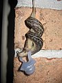
This species has a very unusual and distinctive mating method, where the pair of slugs hang in the air, suspended from a tree branch or other structure, using a thick thread of mucus.
Although native to Europe, this species has been accidentally introduced to many other parts of the world.
Description
External anatomy
The body length of adults of this slug species is 10-20 cm (4-8 in).[3]
The greater part of the body is rounded, but there is a short keel on its tail, with about 48 longitudinal rows of elongate, detached tubercles.[4]
The animal's body color is pale-grey, ash-colored, brownish or sometimes yellowish-white. The body is longitudinally streaked or spotted with black. The shield is always black-spotted. The sole of the foot is ash or yellowish-ash colored, and the color is always uniform.[5] The foot-fringe is pale, with a row of minute submarginal blackish tubercles.[4]
Every Limax maximus differs slightly in its pattern of black spots:
-
 Limax maximus in Hamburg, Germany
 -
Limax maximus actively crawling on a sidewalk in Columbus, Ohio, USA
-
 Limax maximus in Switzerland


The tentacles are very long and slender.[4] The reproductive pore is near the base of the right upper tentacle.[6]
The shield is oblong, about one-third of the total length of the animal. The shield is rounded in front, angular behind, and forming an angle of about 80 degrees when in motion, usually of a similar tint to the body, but boldly marbled or maculate with black, somewhat concentrically and interruptedly ridged around a sub-posterior nucleus.[4]
The pneumostome is just posterior to the mid-point of the mantle, as it is in all Limacidae.
The mucus is colorless and iridescent, and not very adhesive.[4]
Although color varieties have no actual taxonomic significance, a large number of color varieties have been described, prominent among them being the varieties serpentinus, vulgaris, cellarius (typical), johnstoni, maculatus, ferrussaci, obscurus, fasciatus and rufescens, of Alfred Moquin-Tandon, and cornaliee, of Pini.[5][7]
As well as different tints and markings, various shades of color exist in Limax maximus:
-
 A lighter specimen
 -
 A darker specimen


Internal anatomy

HG = hermaphordite gland = ovotestis
HD = hermaphrodite duct
AG = albumen gland
SO = spermoviduct
OV = oviduct
VD = vas deferens = sperm-duct
RS = receptaculum seminis
P = penis
PRM = penis retractor muscle
G = genital pore
The shell of Limax maximus is reduced and internal, under the shield. The occurrence of this internal shell was known to Pliny the Elder; the shell was used by the ancient physicians for the sake of its carbonate of lime.[8]


The calcitic shell is situated beneath the hinder part of the shield, and is perceptible through the skin. The color of the shell is whitish. The shape of the shell is oblong-oval and thin, slightly convex above, and correspondingly concave beneath, with a membranous margin. The apex or nucleus is at the posterior margin but inclined towards the left side, and forming the apophysis by which the shell is organically attached to the animal.[4] The length of the shell is 13 mm (1/2 inch) and the width of the shell is 7 mm (1/4 inch).[4] Shells of different Limacidae species are undiagnostic: in other words, they are not helpful for identification purpose.
Digestive system: The formula of the radula is: 62-73/ × 138-157.[2] The intestine has six convolutions and is without a caecum.[6] Of the six convolutions of the intestine, four are imbedded in the liver, and two hang freely in the body cavity.[6]
The nervous system is composed of the typical ganglia. The pedal ganglia are placed beneath the radula sac and joined together by an anterior and a posterior commissure. The abdominal ganglion lies a little to the right of the median line. The visceral ganglia occupy the angle between the lingual sheath and the oesophagus and the buccal ganglia are widely separated but joined together by a commissure nearly as thick as the ganglia themselves.[4]
Reproductive system: The hermaphrodite gland (HG) is elongated and large, and is connected with spermoviduct (SD) by means of the hermaphrodite duct (HD) which takes its course through a portion of the albumen gland (AG). The spermoviduct is thick and well convoluted, and separates further down into a vas deferens or sperm-duct (VD) and an oviduct (OV). The former opens into the upper end of a very long penis (P), to which a strong retractor muscle (PRM) is attached. The lower portion of the penis unites with that of the oviduct at the genital orifice, so that there is no vestibule. The receptaculum seminis (RS) opens into the lower end of the penis near the junction of the two ducts.[6]
Distribution
Fossil distribution
The internal shells of the different species of Limacidae are not recognizable to the species level. Therefore, the fossil distribution of Limax maximus (and other Limacidae species) is unknown. Unidentified calcitic shells of Limacidae are known from European Tertiary and Quaternary deposits.
Indigenous distribution
This species is widely distributed but it is generally considered to be native to Europe.
The indigenous distribution of Limax maximus is western and southern Europe and part of western Africa also,[2] maybe originally Mediterranean.[9]
Within Europe, this species is known to occur in a number of different countries and islands including:
- Great Britain[10] - Limax maximus was described from a scientific point from England in Animalium Angliae Tres Tractatus … Alter de Cochleis tum Terrestribus tum Fluviatilibus by Dr. Martin Lister among first three British slugs in 1678. However the existence of Limax maximus in England was indicated twelve years before in Christopher Merret's Pinax Rerum Naturalium Britannicarum in 1667.[4]
- Ireland
- Benelux: Belgium, Netherlands[11], Luxembourg
- France
- Switzerland
- Bulgaria
- Scandinavia
- and other areas

Nonindigenous distribution
The non-indigenous distribution of Limax maximus includes:
Central Europe[2]:
Northern Europe - Scandinavia[2]
Eastern Europe[2]:
This slug species has been introduced to North America. It occurs along the East and the West Coasts of that continent.
Its introduction into the United States was first announced by George Washington Tryon in 1867, when it was discovered in cellars in Philadelphia. Within a few years its presence was noticed at Newport, Rhode Island, Brooklyn, Pittsburgh, etc, and it has now become rather numerous in some localities.[5]
East Coast of the United States:
West Coast of the United States:
- California since 1896[12]
- Montana[13]
- Oregon
Other non-indigenous distribution areas include Madeira,[5] southern Africa, Australia, Tasmania, New Zealand[2] and southern Brazil.[14]
Behavior
Limax maximus is a nocturnal animal which feeds at night.[8]
It is inactive in its habits, not very prolific, and exudes a thick and glutinous slime which is iridescent when dried.[8] When alarmed, or at rest, this slug merely draws its head within the shield, but does not otherwise contract its body. When irritated, it is said to expand its shield.[8]
The homing faculty is strongly developed in this species, which, after its nocturnal rambles or foraging expeditions, usually returns to the particular crevice or chink in which it has established itself.[4]
Limax maximus is capable of associative learning, specifically classical conditioning, because it is capable of aversion learning and other types of learning.[15][16] They can also detect that there are deficiencies in a nutritionally incomplete diet, if the essential amino acid methionine is experimentally removed from their food.[17]
Ecology
Habitat
The slugs are almost always found near human habitation — usually in lawns, gardens, cellars or in other damp areas.
This species is not gregarious. It frequents gardens, damp and shady hedgerows and woods, hiding during the day beneath stones, under fallen trees, or other obscure and damp places. It does however exhibit a decided preference for the vicinity of human habitations, and readily takes up its abode in damp cellars or outbuildings.[4]
In Ireland, this predilection for human dwellings is not exhibited, and the species is restricted to woods and other similar places. It may even be met with almost within high-water mark on the seashore.[4]
Feeding habits
Limax maximus is omnivorous and feeds on plants and on fungi.[4] It can also be carnivorous, feeding on other slugs. It can move four times faster than the banana slug[18], which is an herbivorous slug of comparable size.
Life cycle

The eggs of this slug are deposited in a cluster, slightly attached to each other.[8] Eggs are transparent, elastic and slightly yellowish in color.[6] The size of the egg is 6×4.5 mm.[19][20] They hatch in about a month.[4]
The tiny slugs which emerge from the eggs need at least two years to reach sexual maturity.[21]
The lifespan of Limax maximus is 2.5–3 years.[22]
Mating
The mating habits of Limax maximus are considered unusual among slugs: the hermaphrodite slugs court, usually for hours, by circling and licking each other. After this, the slugs will climb into a tree or other high area and then, entwined together, lower themselves on a thick string of mucus, evert their white translucent mating organs (penises) from their gonopores (openings on the right side of the head), entwine these organs, and exchange sperm. Both participants will later lay hundreds of eggs.
A commonly seen practice among many slugs is apophallation, when one or both of the slugs chews off the other's penis. The penis of these species is curled like a corkscrew and often becomes entangled in their mate's genitalia in the process of exchanging sperm. When all else fails, apophallation allows the slugs to separate themselves. Once its penis has been removed, a slug is still able to participate in mating subsequently, but only using the female parts of its reproductive system.
-
 Two mating Limax maximus suspended by a mucus thread (drawing 1 below)
 -
 Mating Limax maximus (drawing 6 below)
 -
 Close up of mating Limax maximus (drawing 6 below)
 -
 Close up of mating Limax maximus (drawing 8 below)


1- penises after protrusion from the body. 2 - commencement of the appearance of the frill. 3 - frill partially unroled. 4 - frill completelly expanded, preparatory to twisting together. 5 - penises tightly coiled together, formin the whorled knot. 6 - the succeeding umbrella form. 7 - umbrella form with horizontal margins reversed. 8 - umbrella form with double margins.
Parasites
Parasites of Limax maximus include:
- The nematode worm Agfa flexilis (Dujardin, 1845) lives in the slug's salivary glands[4][unreliable source?]
- The nematode Angiostoma limacis Dujardin, 1845 lives in the rectum[4][unreliable source?]
- Angiostrongylus costaricensis Morera & Cespedes, 1971[14]
This slug is often infested (as are some other slug species) by a white parasitic slug mite Riccardoella limacum. These mites can be seen swarming about its body, and live in its respiratory cavity.[8]
Angiostrongylus cantonensis
A meningitis-causing nematode, Angiostrongylus cantonensis, which normally infests the lungs of rats, has a larval stage which can only live in mollusks, including slugs.
Previously, this nematode was only known to be a problem in tropical areas, but it has since spread to other regions. Live slugs that are accidentally eaten with improperly cleaned vegetables (such as lettuce), or improperly cooked slugs (for use in recipes requiring larger slugs such as banana slugs), can act as a vector for a parasitic infection.[23][24]
References
This article incorporates public domain text from references [4][5][6][8] .
- ^ Template:La icon Linnaeus C. 1758. Systema naturae per regna tria naturae, secundum classes, ordines, genera, species, cum characteribus, differentiis, synonymis, locis. Tomus I. Editio decima, reformata. pp. [1-4], 1-824. Holmiae. (Salvius).
- ^ a b c d e f g h Template:Pl icon Wiktor A. 1989. Limacoidea et Zonitoidea nuda. Slimaki pomrowioksztaltne (Gastropoda: Stylommatophora). Fauna Poloniae 12, Polska Akademia Nauk, Warszawa, 208 pp., 165-168.
- ^ Template:De icon M.P. Kerney, A. D. Cameron, J. H. Jungbluth: Die Landschnecken Nord- und Mitteleuropas. Verlag Paul Parey, Hamburg und Berlin 1983, ISBN 3-490-17918-8, page 183.
- ^ a b c d e f g h i j k l m n o p q Taylor J. W. (7 November) 1902. part 8, pages 1-52. Monograph of the land and freshwater Mollusca of the British Isles. Testacellidae. Limacidae. Arionidae. Taylor Brothers, Leeds. Introduction page XV., pages 34-52.
- ^ a b c d e Tryon G. W. 1885. Manual of Conchology. Second series: Pulmonata Volume 1. Testacellidae, Oleacinidae, Streptaxidae, Helicoidea, Vitrinidae, Limacidae, Arionidae. 364 pp., 60 plates, pages 189-190, plate 46 figure 31-35, 39; plate 49, figure 76.
- ^ a b c d e f Scharff R. F. (July) 1891. The slugs of Ireland. The Scientific Transactions of the Royal Dublin Society, volume IV., series II. Dublin, Royal Dublin Society; London, Williams & Norgate. 513-563. Limax maximus on pages page 517-521. plate LVII.
- ^ Pini. 1876. Bull. Soc. Mal. Ital. ii., 83. (There is described Limax cornaliae Pini)
- ^ a b c d e f g Jeffreys J. G. 1862. British conchology: or, an account of the Mollusca which now inhabit the British Isles and the surrounding seas. Volume I. Land and freshwater shells. page 137-138.
- ^ Template:Sk icon Lisický M. J. 1991. Mollusca Slovenska [The Slovak molluscs]. VEDA vydavateľstvo Slovenskej akadémie vied, Bratislava, 344 pp.
- ^ Michael P. Kerney & R.A.D. Cameron, 1979, A field guide to the land snails of Britain and north-west Europe, Collins, Glasgow.
- ^ Template:Nl icon Limax maximus. Stichting ANEMOON, accessed 10 August 2009
- ^ Vanatta E. G.(June) 1904. Addtionial Notes on Limax maximus L. in California The Nautilus, volume 18, number 2, page 23.
- ^ Giant Gardenslug — Limax maximus. Montana Field Guide. Retrieved on 31 July 2009.
- ^ a b Teixeira C.G., Thiengo S.C., Thome J.W., Medeiros A.B., Camillo-Coura L. & Agostini A.A. 1993. On the diversity of mollusc intermediate hosts of Angiostrongylus costaricensis Morera & Cespedes, 1971 in southern Brazil. Mem Inst Oswaldo Cruz. 1993 Jul-Sep;88(3):487-9. (abstract)
- ^ Sahley C., Rudy J. W. & Gelperin A. (March) 1981. An analysis of associative learning in a terrestrial mollusc . Journal of Comparative Physiology A: Neuroethology, Sensory, Neural, and Behavioral Physiology. Volume 144 (1): 1-8. DOI 10.1007/BF00612791.
- ^ Sahley C.L., Martin K.A. & Gelperin A. 1990. Analysis of associative learning in the terrestrial mollusc Limax maximus. II. Appetitive learning. J Comp Physiol A. 1990 Aug;167(3):339-45.
- ^ Delaney K. & Gelperin A. (September) 1986. http://www.springerlink.com/content/r2h118662466738m/. Journal of Comparative Physiology. A, Sensory, neural, and behavioral physiology 159(3):281-95.
- ^ Rosetta R. 2007. Great Gray Garden Slug, Tiger slug, spotted leopard slug. Last edited 15 October 2007, accessed 1 August 2009.
- ^ Heller J.: Life History Strategies. in Barker G. M. (ed.): The biology of terrestrial molluscs. CABI Publishing, Oxon, UK, 2001, ISBN 0-85199-318-4. 1-146, cited page: 428.
- ^ Template:De icon Frömming E. 1954. Biologie der Mitteleuropäischen Landgastropoden. Duncker & Humblot, Berlin.
- ^ Limax maximus, Invertebrate Anatomy OnLine, Lander University.
- ^ Limax maximus . UF / IFAS Featured Creatures Web site, publication date: June 1999, latest revision: May 2009. accessed 1 August 2009.
- ^ Sanjaya N. Senanayake, Don S. Pryor, John Walker & Pam Konecny 2003. First report of human angiostrongyliasis acquired in Sydney. The Medical Journal of Australia, 179 (8): 430-431.
- ^ Anna Salleh (20 October 2003). "Man's brain infected by eating slugs".
Further reading
- Gelperin A. 1974. Olfactory Basis of Homing Behavior in the Giant Garden Slug, Limax maximus. Proc Natl Acad Sci U S A. 1974 March; 71(3): 966–970.
- Wieland S. J. & Gelperin A. 1983. Dopamine elicits feeding motor program in Limax maximus. J Neurosci 1983, 3:1735-1745.