Lysosome: Difference between revisions
Content deleted Content added
m Reverted edits by 165.138.80.81 (talk) to last revision by ClueBot NG (HG) |
←Replaced content with '{{Organelle diagram}} '''Lysosomes''' are cells in your body.' |
||
| Line 1: | Line 1: | ||
{{Organelle diagram}} |
{{Organelle diagram}} |
||
'''Lysosomes''' are cells in your body. |
|||
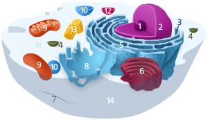
'''Lysosomes''' are cellular [[organelle]]s that contain acid [[hydrolase]] enzymes that break down waste materials and cellular debris. These are non-specific. They can be described as the stomach of the cell. They are found in animal cells, while their existence in yeasts and plants are disputed. Some biologists say the same roles are performed by lytic [[vacuole]]s,<ref>{{cite journal |author=Samaj J, Read ND, Volkmann D, Menzel D, Baluska F |title=The endocytic network in plants |journal=Trends Cell Biol. |volume=15 |issue=8 |pages=425–33 |year=2005 |month=August |pmid=16006126 |doi=10.1016/j.tcb.2005.06.006}}</ref> while others suggest there is strong evidence that lysosomes are indeed in some plant cells.<ref>{{cite journal |author=Sarah J. Swansona, Paul C. Bethkea, and Russell L. Jonesa|title=Barley Aleurone Cells Contain Two Types of Vacuoles: Characterization of Lytic Organelles by Use of Fluorescent Probes|journal=The Plant Cell|volume=10 |issue=5 |pages=685–689 |year=1998 |month=May }}</ref> Lysosomes digest excess or worn-out [[organelle]]s, food particles, and engulf [[virus]]es or [[bacteria]]. The [[biological membrane|membrane]] around a lysosome allows the [[digestive enzyme]]s to work at the 4.5 [[pH]] they require. Lysosomes fuse with [[vacuole]]s and dispense their enzymes into the [[vacuole]]s, digesting their contents. They are created by the addition of hydrolytic enzymes to early [[endosome]]s from the [[Golgi apparatus]]. The name ''lysosome'' derives from the Greek words '''[[lysis]]''', ''to separate'', and '''soma''', ''body''. They are frequently nicknamed "suicide-bags" or "suicide-sacs" by cell biologists due to their [[autolysis]]. Lysosomes were discovered by the Belgian cytologist [[Christian de Duve]] in the 1960s. |
|||
The size of a lysosome varies from 0.1–1.2 [[micrometre|μm]].<ref>{{cite book | last=Kuehnel | first=W | title=Color Atlas of Cytology, Histology, & Microscopic Anatomy | publisher=Thieme | year=2003 | edition=4th | pages=34 | isbn=1-58890-175-0 }}</ref> At [[pH]] 4.8, the interior of the lysosomes is acidic compared to the slightly alkaline [[cytosol]] (pH 7.2). The lysosome maintains this pH differential by pumping [[proton]]s (H<sup>+</sup> ions) from the cytosol across the [[Cell membrane|membrane]] via [[proton pump]]s and chloride [[ion channel]]s. The lysosomal membrane protects the cytosol, and therefore the rest of the [[cell (biology)|cell]], from the [[degradative enzyme]]s within the lysosome. The cell is additionally protected from any lysosomal acid [[hydrolases]] that drain into the cytosol, as these enzymes are pH-sensitive and do not function well or at all in the alkaline environment of the cytosol.This ensures that cytosolic molecules and organelles are not lysed in case there is leakage of the hydrolytic enzymes from the lysosome. |
|||
'''Lysosomes''' are the cell's waste disposal system and [[Residual body|can digest some compounds]]. They are used for the digestion of [[macromolecule]]s from [[phagocytosis]] (ingestion of other dying cells or larger extracellular material, like foreign invading microbes), [[endocytosis]] (where [[Receptor (biochemistry)|receptor protein]]s are recycled from the cell surface), and [[autophagy]] (where in old or unneeded organelles or proteins, or microbes that have invaded the cytoplasm are delivered to the lysosome). Autophagy may also lead to [[autophagy|autophagic cell death]], a form of [[programmed cell death|programmed self-destruction]], or [[autolysis]], of the cell, which means that the cell is digesting itself. |
|||
Other functions include digesting foreign bacteria (or other forms of waste) that invade a cell and helping repair damage to the [[plasma membrane]] by serving as a membrane patch, sealing the wound. In the past, lysosomes were thought to kill cells that are no longer wanted, such as those in the tails of [[tadpole]]s or in the web from the fingers of a 3- to 6-month-old [[fetus]]. |
|||
== Formation == |
|||
Many components of the animal cell can be seen recycling and sharing their membranous units. For instance, in [[endocytosis]], a portion of the cell’s plasma membrane pinches off to form a vesicle that will eventually fuse with an organelle within the cell. Without active replenishment, the plasma membrane would continuously decrease in size. It is thought that lysosomes participate in this dynamic membrane exchange system and are formed by a gradual maturation process from [[endosomes]].<ref name=Alberts>{{cite book|last=Alberts|first=Bruce et al.|title=Molecular biology of the cell|year=2002|publisher=Garland Science|location=New York|isbn=0-8153-3218-1|edition=4th}}</ref> |
|||
The production and transport of lysosomal proteins suggest one method of lysosome sustainment. Lysosomal protein genes are transcribed in the [[nucleus]]. mRNA transcripts exit the nucleus into the cytosol, where they are translated by [[ribosomes]]. The nascent peptide chains are [[Protein targeting#Protein translocation|translocated]] into the rough [[endoplasmic reticulum]], where they are modified. Upon exiting the endoplasmic reticulum and entering the [[Golgi apparatus]] via vesicular transport, a specific lysosomal tag, [[mannose 6-phosphate]], is added to the peptides. The presence of these tags allow for binding to mannose 6-phosphate receptors in the golgi apparatus, a phenomenon that is crucial for proper packaging into vesicles destined for the lysosomal system.<ref name=Lodish>{{cite book|last=Lodish|first=Harvey et al.|title=Molecular cell biology|year=2000|publisher=Scientific American Books|location=New York|isbn=0-7167-3136-3|edition=4th}}</ref> |
|||
Upon leaving the golgi apparatus, the lysosomal enzyme-filled vesicle fuses with a late endosome, a relatively acidic organelle with an approximate pH of 5.5.<ref name=Lodish /> This acidic environment causes dissociation of the lysosomal enzymes from the mannose 6-phosphate receptors<ref name=Lodish />. The enzymes are packed into vesicles for further transport to established lysosomes.<ref name=Lodish /> The late endosome itself can eventually mature into a mature lysosomes, as evidenced by the transport of endosomal membrane components from the lysosomes back to the endosomes.<ref name=Alberts /> |
|||
== Lysosomotropism == |
|||
Weak [[Base (chemistry)|bases]] with [[Lipophilicity|lipophilic]] properties accumulate in acidic intracellular compartments like lysosomes. While the plasma and lysosomal membranes are permeable for neutral and uncharged species of weak bases, the charged protonated species of weak bases do not permeate biomembranes and accumulate within lysosomes. The concentration within lysosomes may reach levels 100 to 1000 fold higher than extracellular concentrations. This phenomenon is called "lysosomotropism"<ref>de Duve C, de Barsy T, Poole B, Trouet A, Tulkens P, van Hoof F. Lysosomotropic agents. Biochem.Pharmacol. 23:2495-2531, 1974. PMID 4606365</ref> or "acid trapping". The amount of accumulation of lysosomotropic compounds may be estimated using a cell based mathematical model.<ref>Trapp S, Rosania G, Horobin RW, Kornhuber J. Quantitative modeling of selective lysosomal targeting for drug design. Eur.Biophys.J. 37 (8):1317-1328, 2008. PMID 18504571</ref> |
|||
A significant part of the clinically approved drugs are lipophilic weak bases with lysosomotropic properties. This explaines a number of pharmacological properties of these drugs, such as high tissue-to-blood concentration gradients or long tissue elimination half-lifes; these properties have been found for drugs such as [[haloperidol]],<ref>Kornhuber J, Schultz A, Wiltfang J, Meineke I, Gleiter CH, Zöchling R, Boissl KW, Leblhuber F, Riederer P. Persistence of haloperidol in human brain tissue. Am.J.Psychiatry 156:885-890, 1999. PMID 10360127</ref> [[levomepromazine]] <ref>Kornhuber J, Weigmann H, Röhrich J, Wiltfang J, Bleich S, Meineke I, Zöchling R, Hartter S, Riederer P, Hiemke C. Region specific distribution of levomepromazine in the human brain. J.Neural Transm. 113:387-397, 2006. PMID 15997416</ref> and [[amantadine]].<ref>Kornhuber J, Quack G, Danysz W, Jellinger K, Danielczyk W, Gsell W, Riederer P. Therapeutic brain concentration of the NMDA receptor antagonist amantadine. Neuropharmacology 34:713-721, 1995. PMID 8532138</ref> However, in addition to high tissue concentrations and long elimination half-life is explained also by lipophilicity and absorption of drugs to fatty tissue structures. Important lysosomal enzymes, such as acid sphingomyelinase, may be inhibited by lysososomally accumulated drugs.<ref>Kornhuber J, Tripal P, Reichel M, Terfloth L, Bleich S, Wiltfang J, Gulbins E. Identification of new functional inhibitors of acid sphingomyelinase using a structure-property-activity relation model. J.Med.Chem. 51:219-237, 2008. PMID 18027916</ref><ref>Kornhuber J, Muehlbacher M, Trapp S, Pechmann S, Friedl A, Reichel M, Mühle C, Terfloth L, Groemer TW, Spitzer GM, Liedl KR, Gulbins E, Tripal P. Identification of novel functional inhibitors of acid sphingomyelinase. PLoS ONE 6 (8):e23852, 2011. PMID 21909365</ref> Such compounds are termed FIASMAs (functional inhibitor of acid sphingomyelinase)<ref>Kornhuber J, Tripal P, Reichel M, Mühle C, Rhein C, Muehlbacher M, Groemer TW, Gulbins E. Functional inhibitors of acid sphingomyelinase (FIASMAs): a novel pharmacological group of drugs with broad clinical applications. Cell.Physiol.Biochem. 26:9-20, 2010. PMID 20502000</ref> and include for example [[fluoxetine]], [[sertraline]] or [[amitriptyline]]. |
|||
==References== |
|||
<references /> |
|||
==External links== |
|||
{{wiktionary}} |
|||
* {{NCBI-scienceprimer}} |
|||
* [http://opm.phar.umich.edu/localization.php?localization=Lysosome%20membrane 3D structures of proteins associated with lysosome membrane] |
|||
* [http://www.hideandseek.org Hide and Seek Foundation For Lysosomal Research Team] |
|||
* [http://www.nytimes.com/2009/10/06/science/06cell.html Self-Destructive Behavior in Cells May Hold Key to a Longer Life] |
|||
* [http://content.nejm.org/cgi/content/full/NEJMoa0902630 Mutations in the Lysosomal Enzyme–Targeting Pathway and Persistent Stuttering] |
|||
* [http://highered.mcgraw-hill.com/olc/dl/120067/bio01.swf Animation showing how Lysosomes are made, and their function] |
|||
{{organelles}} |
|||
[[Category:Organelles]] |
|||
[[af:Lisosoom]] |
|||
[[ar:يحلول]] |
|||
[[az:Lizosomlar]] |
|||
[[be:Лізасома]] |
|||
[[bg:Лизозома]] |
|||
[[bs:Lizozom]] |
|||
[[ca:Lisosoma]] |
|||
[[cs:Lyzozom]] |
|||
[[da:Lysosom]] |
|||
[[de:Lysosom]] |
|||
[[et:Lüsosoom]] |
|||
[[el:Λυσόσωμα]] |
|||
[[es:Lisosoma]] |
|||
[[eo:Lizosomo]] |
|||
[[eu:Lisosoma]] |
|||
[[fa:کافندهتن]] |
|||
[[fr:Lysosome]] |
|||
[[gv:Leesosoom]] |
|||
[[gl:Lisosoma]] |
|||
[[ko:리소좀]] |
|||
[[hi:लाइसोसोम]] |
|||
[[hr:Lizosom]] |
|||
[[id:Lisosom]] |
|||
[[it:Lisosoma]] |
|||
[[he:ליזוזום]] |
|||
[[jv:Lisosom]] |
|||
[[ka:ლიზოსომა]] |
|||
[[kk:Лизосома]] |
|||
[[la:Lysosoma]] |
|||
[[lv:Lizosoma]] |
|||
[[lb:Lysosom]] |
|||
[[lt:Lizosomos]] |
|||
[[hu:Lizoszóma]] |
|||
[[mk:Лизозом]] |
|||
[[ml:ലൈസോസോം]] |
|||
[[nl:Lysosoom]] |
|||
[[ja:リソソーム]] |
|||
[[no:Lysosom]] |
|||
[[oc:Lisosòma]] |
|||
[[pl:Lizosom]] |
|||
[[pt:Lisossomo]] |
|||
[[ro:Lizozom]] |
|||
[[ru:Лизосома]] |
|||
[[si:ලයිසොසෝම]] |
|||
[[simple:Lysosome]] |
|||
[[sk:Lyzozóm]] |
|||
[[sl:Lizosom]] |
|||
[[ckb:لایسۆسۆم]] |
|||
[[sr:Лизозом]] |
|||
[[sh:Lizozom]] |
|||
[[su:Lisosom]] |
|||
[[fi:Lysosomi]] |
|||
[[sv:Lysosom]] |
|||
[[ta:இலைசோசோம்]] |
|||
[[th:ไลโซโซม]] |
|||
[[tr:Lizozom]] |
|||
[[uk:Лізосома]] |
|||
[[vi:Tiêu thể]] |
|||
[[zh:溶酶体]] |
|||
Revision as of 18:52, 12 November 2012
| Cell biology | |
|---|---|
| Animal cell diagram | |
 Components of a typical animal cell:
|
Lysosomes are cells in your body.
