
List of instruments used in ophthalmology
(Redirected from Strabismus hook)
This is a list of instruments used in ophthalmology.[1]
Instrument list[edit]
A complete list of ophthalmic instruments can be found below:
| Instrument | Uses |
|---|---|
| Toric Marker | to mark 0 to 180 degree reference mark for Toric IOL implant |
| Pre-chopper | to chop lens into pieces before implantation new lens and reduce phaco time |
| Spectacles (glasses) | to correct refractive errors of the eye; not invasive |
| Contact lenses | to correct refractive errors of the eye; a little invasive |
| Phoropter | used in refraction testing |
| Tonometers | used to determine the intraoccular pressure (IOP) - useful in glaucoma; video link for various types of tonometers. |
| Speculum: | to keep the eyes open during any operation |
| Universal eye speculum | -do-; heavy instrument and can not keep eyelashes out of the operating field |
| •Guarded eye speculum (left and right) | -do-; heavy instrument but can keep eyelashes out of the operating field with its "guard" and hence left or right ones are required |
| •Wire Speculum | to keep the eyes open during any operation; light wire instrument |
| Needle holders: | holding the needle in position while applying sutures |
| •Silcock's needle holder | -do-; has a catch and is used for heavier gauge needles; used mainly for skin, muscle and corneal incisions |
| •Arruga's needle holder | -do-; has a catch (lock) and is used for heavier gauge needles (thicker than 6–0); used mainly for skin, muscle and corneal incisions |
| •Barraquer's needle holder | -do-; small instrument with a spring action with or without a catch used for finer gauge needles (5-0 or finer); used mainly for intraoccular incisions |
| Forceps: | to hold anything |
| •Artery forceps (haemostat) | medium-sized, with a serrated tip and a catch; used to hold bleeding vessels and compress them in order to make them stop bleeding and also to hold or crush structures. |
| •Fixation forceps | has a few teeth at the tip; for holding structures and restricting their movement or to hold small swabs |
| •Plain dissecting forceps | blunt untoothed with a serrated tip; for holding structures and restricting their movement or to hold small swabs |
| •Iris forceps | fine tipped (straight or otherwise) with small teeth; to hold the iris tissue during procedures |
| •Elschnig's intracapsular forceps | fine untoothed forceps for holding tissue, swabs, sutures, etc.; removing things like clots, capsule fragments, lens, etc.; used in cataract surgery |
| •Arruga's intracapsular forceps | fine untoothed forceps holding tissue, swabs, sutures, etc.; removing things like clots, capsule fragments, lens, etc.; used in cataract surgery |
| •Colibri forceps | fine toothed forceps for holding flaps of cornea or sclera and rarely the iris |
| •Saint Martin's forceps | holding flaps of cornea or sclera and rarely the iris |
| •Superior rectus holding forceps | specially curved (to fit into the orbit of the eye) forceps for catching hold of the muscle bellies of the intraorbital muscles and sutures |
| •Suture tier forceps | fine limbed untoothed forceps to hold fine sutures or hairs |
| •Capsulotomy forceps | to tear the anterior capsule of the lens during cataract surgery |
| •Disc holding forceps | used in glaucoma surgery (obsolete) |
| •Capsulorhexis forceps | fine sharp-tipped untoothed forceps for doing a continuous curvilinear incision and removal of the anterior capsule of the lens ("continuous curvilinear capsulorhexis - ccc") |
| •MacPherson's forceps | fine sharp-tipped untoothed forceps with an angulation for holding parts of the lens, the intraocular lens, 10-0 (very fine) sutures, etc. |
| •Chalazion forceps (clamp) | self-retaining with discoid ends; used to hold and prevent a chalazion from bleeding during its surgery |
| Diamond knife | used to perform microincisions on the cornea in the Radial keratotomy and Mini Asymmetric Radial Keratotomy (M.A.R.K.) |
| •Epilation forceps (Cilia forceps) | stout flat-ended blunt forceps with a thickened end to remove eyelashes |
| •Entropion forceps | self-retaining with big discoid ends used to hold and prevent an entropion from bleeding during its surgery |
| Chalazion scoop | to remove the granulation tissue from a chalazion during surgery |
| Entropion clamp | right and left varieties exist; large clamp with two limbs; self-retaining with big discoid ends used to hold and prevent an entropion from bleeding during its surgery |
| Nettleship's punctum dilator | to dilate the lacrimal punctum of the lacrimal apparatus of the eye for syringing or operations |
| Cystotome | a 26 gauge needle bent twice used for incising the anterior capsule of the lens in lens extraction |
| Wire vectis | a loop of wire attached to a stack used to extract cataract affected lenses |
| Irrigating vectis | a small hollow instrument with a used to introduce fluid into the anterior chamber to raise its pressure to aid cataract extraction [2] |
| Canula | used to carry fluid |
| •Irrigation-aspiration two-way canula | effectively two small canulae fitted together, one to introduce fluid and the other to extract the cortical materials, blood, etc. in eye operations |
| •Lacrimal canula | small curved canula the size of a syringe needle used to introduce fluids or drugs into the nasolacrimal passage to test its patency or during surgery (dacrocystography, dacrocystectomy, dacryocystorhinostomy(DCR), etc. |
| Lang's lacrimal dissector with scoop | for blunt dissections and cleaning during operations like dacryocystorhinostomy |
| Rougine | dissection of lacrimal sac |
| Retractor | to pull and hold overlying tissue out of the operating field |
| •Muller's self retaining adjustable haemostatic retractor | -do-; self retaining haemostatic |
| •Cat's paw retractor | -do- |
| •Desmarre's lid retractor | -do-; specially for noncooperative patients and to see the fornices (see human eye) |
| Bone punch | to fracture pieces from a thin bone in facial surgery and during operations like dacryocystorhinostomy |
| Evisceration spoon or scoop | removing all the contents of the eyeball during evisceration (complete removal of all structures within the eye in diseases like endophthalmitis |
| Lid plate | flat large instrument that has a groove and is placed between the lid and globe of the eye to provide a solid support for eyelid surgery |
| Hammer, chisel and bone gouge | bone cutting and shaping |
| Bowmen's discission needle | microsurgery of the lens capsule[3] |
| Knives | to cut structures |
| •Surgical scalpel with small blades | general purpose instrument |
| •von Graefe's cataract knife | cutting out of the anterior chamber from the inside through the limbus |
| •Tookes' knife (Sclero-corneal splitter) | making sclerocorneal tunnels in "small incision cataract surgery (SICS)" and keratoplasty |
| •Crescent knife (Sclero-corneal splitter) | making sclerocorneal tunnels in "small incision cataract surgery" |
| •Angular keratome | making sclerocorneal tunnels in "small incision cataract surgery"; larger one used to increase the size of the incision |
| •Side-port blade | making sclerocorneal "side port" (a secondary tunnel) tunnels in "small incision cataract surgery" |
| •Beer's knife | incise the conjunctiva or the eyelid skin |
| •Keratotome | small triangular blade with two sharp edges used to incise the limbus (sclerocorneal junction) |
| •Zeigler's knife | very tiny knife for intaoccular maneuvers specially when space is less |
| Scissors | - |
| •Conjunctival sac scissors | flat small curved scissors to cut the conjunctive |
| •Corneal spring scissors | medium spring-open used to cut the external side of the cornea, fine sutures; iris, etc. |
| •de' Wecker's iris scissors | small slender spring-open scissors for intraoccular maneuvers (iris and deeper and more delicate structures); has two wings to operate it and one sharp and one blunt blade. |
| •Vannas' scissors | small slender spring-open scissors for intraoccular maneuvers (iris and deeper and more delicate structures); has two wings to operate it and one sharp and one blunt blade. |
| •Enucleation scissors | thick scissors used to cut the optic nerve in enucleation operation |
| Bowman's lacrimal probe | probing the nasolacrimal duct |
| Lens expressor | used to force out the lens in extracapsular or intracapsular cataract extraction |
| McNamar's spoon | used to force out the lens in intracapsular cataract extraction |
| Iris repositor | two limbed instrument used to remove the iris during posterior chamber maneuvers |
| Sinsky's hook intraocular lens dialler | angulated round hook with a handle used in insertion of an intraocular lens |
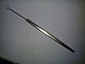
| Strabismus hook | muscle hook or squint hook; sharp tip or knobbed tip; used in squint surgery |
| Foreign body spud and needle | Spud to remove superficial and needle for the deep foreign bodies in the eye |
| Elliot's trephine with handle | used in corneal donation (eye donation) to cut out the cornea in a circular fashion |
| Castroveijo's calipers | various measurements are taken |
| Castroveijo's corneal trephine | used in corneal donation (eye donation) to cut out the cornea in a circular fashion |
| Pin-hole | testing visual acuity |
| Red green goggles | (red - right side & green - left side) used in Worth 4 dot test, diplopia testing |
| Prisms | to measure the degree of squints; in other instruments; refractive correction; etc. |
| Placido's disc | to assess the condition of the corneal surface |
| Retinoscope | objective determination of refractive error and for looking inside the eye |
| Loupe | used to search for magnified examination of the anterior segment of the eye (uniocular or binocular) |
| Jackson's cross cylinder | used to check the power and axis of a cylindrical lens |
| Maddox rod | used to test for latent squint and retinal function |
| Refraction box | has lenses of different powers for refraction testing |
| Slit lamp bio microscope | used for examining the anteriorly placed structures the eye; video link |
| Charts for vision | - |
| •Distant vision | to determine visual acuity of distant vision |
| ••Snellen's distant vision chart | -do-; for those who can read in English |
| ••Regional language charts | -do-; for those who can read in their local language |
| ••E Chart | -do-; for those who can not read |
| ••Landolt's broken ring chart | -do-; for those who can not read |
| ••Toys pr picture chart | -do-; for children |
| •Near vision | -do-; to determine visual acuity of near vision |
| ••Jager's chart | -do- |
| ••Printer's types of N series | -do- |
| ••Snellen's near chart (1/17th reduction of distant chart) | -do-; standard chart of alphabets; video link |
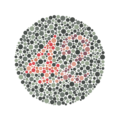
| •Colour vision: | to test colour vision |
| ••Ishihara's chart | to determine the type of colour blindness |
| Stenopaeic slit | detection of axis of the cylindrical (astigmatism) power of the eye; glaucoma testing |
| Implants | - |
| •Intraocular lens | prosthetic lenses implanted after lens (anatomy) removal |
| •Artificial eyes | as non-functional cosmetic implants into the eye socket |
| Blade breaker | to break disposable blade after use to prevent reuse |
| Thermo-cautery | to coagulate blood vessels and prevent haemorrhage |
| Cryoprobe | to freeze and extract the lens |
| Yttrium aluminium garnet laser (YAG laser) | to correct posterior capsular opacification (specially after removal of a cataract, if required), peripheral iridotomy, retinal surgery, laser-assisted sub-epithelial keratectomy (LASEK)[4] etc. |
| Electrolysis | used for permanent hair removal |
| Electrocautery | for electrosurgery |
| Phacoemulsification | used for extraction of a cataract affected lens after emulsifying it using a high frequency (energy) ultrasound probe [5] |
Image gallery[edit]
-
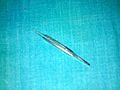
 Akahoshi Combo II Prechopper
 -
 Glasses
 -
 Contact lenses
 -
 Plain dissecting forceps
 -
 Artery forceps or Haemostat
 -
 Mosquito forceps
 -
 Linen holding forceps
 -
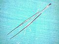
 Bowman's lacrimal probe
 -
 Saint Martin's forceps
 -
 Eye Lens expressor
 -
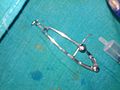
 Nettleship's punctum dilator
 -
 Small scissors
 -
 Scalpel with blade attached
 -
 Conjunctival sac scissors
 -
 Barraquer's needle holder
 -
 Lacrimal sac dilator with scoop
 -
 Muller's retractor, top view
 -
 Muller's retractor, bottom view
 -
 Angular keratotome
 -
 Long dissecting forceps
 -
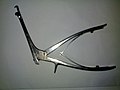
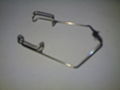
 Universal eye speculum
 -
 Rougine
 -
 Iris repositor
 -
 Irrigating vectis
 -
 Lacrimal dissector with scoop
 -
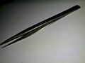
 Special blades
 -
 von Graefe's cataract knife
 -
 Foreign body spud and needle
 -
 Cystitome
 -
 Angular keratotomes
 -
 Barraquer's needle holder
 -
 A bone punch
 -
 Callipers
 -
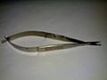 Corneal spring scissors
 -
 Intraoccular lenses in their cases
 -
 Intraoccular lens in place
 -
 Intraoccular lens "dialer" or Sinsky hook
 -
 Irrigating aspirating bi-way cannula
 -
 Lenses used for refraction testing
 -

-
 Suture tying forceps for fine sutures like 8-0
 -
 Upper one: Suture tying forceps; Lower one: Iris forceps; For comparison
 -
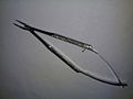
 Upper right: Corneal spring scissors; Lower left: Vanna's scissors; for comparison
 -
 Vanna's scissors
 -
 Wire speculum
 -
 Wire vectis
 -
 Plain dissecting forceps
 -
 Thermocautery
 -
 A standard illuminated E chart
 -
 A standard illuminated Snellen's chart for distant vision
 -
 A set of lenses used in refraction testing
 -
 Ishihara Plate 9
 -
 Ishihara Plate 23
 -
 A phoropter
 -
 NdYAG Laser
 -
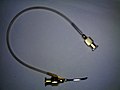
 Lacrimal canula


References[edit]
- ^ Ophthalmology Oral & Practical 3rd edition, by Dr. Danesh ISBN 81-86793-66-6
- ^ Irrigating vectis – Patent 4479802
- ^ Billson FA, Thurgood R, Perriam DJ (December 1975). "Discission needle". Br J Ophthalmol. 59 (12): 741. doi:10.1136/bjo.59.12.741. PMC 1017447. PMID 1218187.
- ^ US FDA/CDRH: LASIK – Learning About LASIK Archived 2004-11-20 at the Wayback Machine
- ^ Untitled Document