Synovial fluid

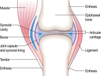
Synovial fluid is a viscous, non-Newtonian fluid found in the cavities of synovial joints. With its yolk-like consistency ("synovial" partially derives from ovum, Latin for egg), the principal role of synovial fluid is to reduce friction between the articular cartilage of synovial joints during movement.
Overview
The inner membrane of synovial joints is called the synovial membrane and secretes synovial fluid into the joint cavity. The fluid contains hyaluronic acid secreted by fibroblast-like cells in the synovial membrane and interstitial fluid filtered from the blood plasma.[1] This fluid forms a thin layer (roughly 50 μm) at the surface of cartilage and also seeps into microcavities and irregularities in the articular cartilage surface, filling all empty space.[2] The fluid in articular cartilage effectively serves as a synovial fluid reserve. During movement, the synovial fluid held in the cartilage is squeezed out mechanically to maintain a layer of fluid on the cartilage surface (so-called weeping lubrication). The functions of the synovial fluid include:
- reduction of friction - synovial fluid lubricates the articulating joints[3]
- shock absorption - as a dilatant fluid, synovial fluid is characterized by the rare quality of becoming more viscous under applied pressure; the synovial fluid in diarthrotic joints becomes thick the moment shear is applied in order to protect the joint and subsequently thins to normal viscosity instantaneously to resume its lubricating function between shocks
- nutrient and waste transportation - the fluid supplies oxygen and nutrients and removes carbon dioxide and metabolic wastes from the chondrocytes within the surrounding cartilage
Composition
Synovial tissue is sterile and composed of vascularized connective tissue that lacks a basement membrane. Two cell types (type A and type B) are present: Type A is derived from blood monocytes, and it removes the wear-and-tear debris from the synovial fluid. Type B produces synovial fluid. Synovial fluid is made of hyaluronic acid and lubricin, proteinases, and collagenases. Synovial fluid exhibits non-Newtonian flow characteristics; the viscosity coefficient is not a constant and the fluid is not linearly viscous. Synovial fluid has thixotropic characteristics; viscosity decreases and the fluid thins over a period of continued stress.[4]
Normal synovial fluid contains 3–4 mg/ml hyaluronan (hyaluronic acid), a polymer of disaccharides composed of D-glucuronic acid and D-N-acetylglucosamine joined by alternating beta-1,4 and beta-1,3 glycosidic bonds.[5] Hyaluronan is synthesized by the synovial membrane and secreted into the joint cavity to increase the viscosity and elasticity of articular cartilages and to lubricate the surfaces between synovium and cartilage.[6]
Synovial fluid contains lubricin secreted by synovial cells. Chiefly, it is responsible for so-called boundary-layer lubrication, which reduces friction between opposing surfaces of cartilage. There also is some evidence that it helps regulate synovial cell growth.[7]
Its functions are:
reducing friction by lubricating the joint, absorbing shocks, and supplying oxygen and nutrients to and removing carbon dioxide and metabolic wastes from the chondrocytes within articular cartilage.[citation needed]
It also contains phagocytic cells that remove microbes and the debris that results from normal wear and tear in the joint.
Health and disease
Collection
Synovial fluid may be collected by syringe in a procedure termed arthrocentesis, also known as joint aspiration.
Classification
Synovial fluid may be classified into normal, noninflammatory, inflammatory, septic, and hemorrhagic:
| Normal | Noninflammatory | Inflammatory | Septic | Hemorrhagic | |
| Volume (ml) | <3.5 | >3.5 | >3.5 | >3.5 | >3.5 |
| Viscosity | High | High | Low | Mixed | Low |
| Clarity | Clear | Clear | Cloudy | Opaque | Mixed |
| Color | Colorless/straw | Straw/yellow | Yellow | Mixed | Red |
| WBC/mm3 | <200 | <2,000[8] | 5,000[8]-75,000 | >50,000[8] | Similar to blood level |
| Polys (%) | <25 | <25[8] | 50[8]-70[8] | >70[8] | Similar to blood level |
| Gram stain | Negative | Negative | Negative | Often positive | Negative |
Pathology

Many synovial fluid types are associated with specific diagnoses:[9][10]
- Noninflammatory (Group I)
- Osteoarthritis, degenerative joint disease
- Trauma
- Rheumatic fever
- Chronic gout or pseudogout
- Scleroderma
- Polymyositis
- Systemic lupus erythematosus
- Erythema nodosum
- Neuropathic arthropathy (with possible hemorrhage)
- Sickle-cell disease
- Hemochromatosis
- Acromegaly
- Amyloidosis
- Inflammatory (Group II)
- Rheumatoid arthritis
- Reactive arthritis
- Psoriatic arthritis
- Acute rheumatic fever
- Acute gout or pseuodgout
- Scleroderma
- Polymyositis
- Systemic lupus erythematosus
- Ankylosing spondylitis
- Inflammatory bowel disease arthritis
- Infection (viral, fungal, bacterial) including Lyme disease
- Acute crystal synovitis
- Septic (Group III)
- Hemorrhagic
- Trauma
- Tumors
- Hemophilia/coagulopathy
- Scurvy
- Ehlers-Danlos syndrome
- Neuropathic arthropathy
Cracking joints
When the two articulating surfaces of a synovial joint are separated from one other, the volume within the joint capsule increases and a negative pressure results. The volume of synovial fluid within the joint is insufficient to fill the expanding volume of the joint and gases dissolved in the synovial fluid (mostly carbon dioxide) are liberated and quickly fill the empty space, leading to the rapid formation of a bubble.[11] This process is known as cavitation. Cavitation in synovial joints results in a high frequency 'cracking' sound.[12][13]
References
- ^ Principles of Anatomy & Physiology, 12th Edition, Tortora & Derrickson, Pub: Wiley & Sons
- ^ http://www.ucl.ac.uk/~regfjxe/NORMALJOINT.htm
- ^ McCracken, Thomas (2000). New Atlas of Human Anatomy. China: Metro Books. pp. 1-240. ISBN 1-58663-097-0.
- ^ "Viscosity". the physics hypertextbook.
- ^ GlycoForum / Science of Hyaluronan-1
- ^ Arthritis - UW Medicine - Department of Orthopaedics and Sports Medicine
- ^ Arthritis Research & Therapy | Full text | Delineating biologic pathways involved in skeletal growth and homeostasis through the study of rare Mendelian diseases that affect bones and joints
- ^ a b c d e f g Table 6-6 in: Elizabeth D Agabegi; Agabegi, Steven S. (2008). Step-Up to Medicine (Step-Up Series). Hagerstwon, MD: Lippincott Williams & Wilkins. ISBN 0-7817-7153-6.
{{cite book}}: CS1 maint: multiple names: authors list (link) - ^ Lupus Anticoagulant
- ^ American College of Rheumatology
- ^ Unsworth A, Dowson D, Wright V. (1971). "'Cracking joints'. A bioengineering study of cavitation in the metacarpophalangeal joint". Ann Rheum Dis. 30 (4): 348–58. doi:10.1136/ard.30.4.348. PMC 1005793. PMID 5557778.
{{cite journal}}: CS1 maint: multiple names: authors list (link) - ^ Watson P, Kernoham WG, Mollan RAB. A study of the cracking sounds from the metacarpophalangeal joint. Proceedings of the Institute of Mechanical Engineering [H] 1989;203:109-118.
- ^ Howstuffworks "What makes your knuckles pop?"
Further reading
- Warman W. "Delineating biologic pathways involved in skeletal growth and homeostasis through the study of rare Mendelian diseases that affect bones and joints." Arthritis Res. Ther. 2003, 5(Suppl 3):5 [1]
