Head louse: Difference between revisions
→Egg and Nit Morphology: replaced secondary source with primary |
Ursasapien (talk | contribs) |
||
| Line 35: | Line 35: | ||
[[Image:Fig.3.Louse egg.jpg|thumb|upright|Louse egg]] |
[[Image:Fig.3.Louse egg.jpg|thumb|upright|Louse egg]] |
||
Louse [[Egg (biology)|eggs]] contain a single embyro, and are attached near the base of a host hair shaft.<ref name="pmid11331679"/> After hatching, the louse [[Nymph_(biology)|nymph]] leaves behind its egg shell, still attached to the hair shaft. |
|||
Louse [[Egg (biology)|eggs]] contain a single embyro, and are attached near the base of a host hair shaft.<ref name="pmid11331679">{{cite journal |author=Williams LK, Reichert A, MacKenzie WR, Hightower AW, Blake PA |title=Lice, nits, and school policy |journal=Pediatrics |volume=107 |issue=5 |pages=1011–5 |year=2001 |pmid=11331679 |doi=}}</ref> To attach the egg, the adult female secretes a quick-hardening glue composed primarily of amino acids.<ref name="pmid10386454">{{cite journal |author=Burkhart CN, Stankiewicz BA, Pchalek I, Kruge MA, Burkhart CG |title=Molecular composition of the louse sheath |journal=J. Parasitol. |volume=85 |issue=3 |pages=559–61 |year=1999 |pmid=10386454 |doi=}}</ref> Each egg is oval-shaped and ca. 0.8 mm in length. Immediately after [[oviposition]], the eggs are shiny, round, and transparent. Viable louse eggs hatch in 6 to 9 days,<ref name="pmid11331679">{{cite journal |author=Williams LK, Reichert A, MacKenzie WR, Hightower AW, Blake PA |title=Lice, nits, and school policy |journal=Pediatrics |volume=107 |issue=5 |pages=1011–5 |year=2001 |pmid=11331679 |doi=}}</ref> but the viability of any particular egg is less than 50%.<ref name="pmid14651472">{{cite journal |author=Burgess IF |title=Human lice and their control |journal=Annu. Rev. Entomol. |volume=49 |issue= |pages=457–81 |year=2004 |pmid=14651472 |doi=10.1146/annurev.ento.49.061802.123253}}</ref> After hatching, the louse [[Nymph_(biology)|nymph]] leaves behind its egg shell, still attached to the hair shaft. |
|||
The term "nit" most correctly refers to the remnants of a hatched egg.<ref name="pmid14651472">{{cite journal |author=Burgess IF |title=Human lice and their control |journal=Annu. Rev. Entomol. |volume=49 |issue= |pages=457–81 |year=2004 |pmid=14651472 |doi=10.1146/annurev.ento.49.061802.123253}}</ref> However, "nit" is used variously to refer to a louse egg (regardless of whether it has hatched or not),<ref name="pmid11331679">{{cite journal |author=Williams LK, Reichert A, MacKenzie WR, Hightower AW, Blake PA |title=Lice, nits, and school policy |journal=Pediatrics |volume=107 |issue=5 |pages=1011–5 |year=2001 |pmid=11331679 |doi=}}</ref> and/or a non-viable (dead) louse egg.<ref name="pmid16911370"/> |
The term "nit" most correctly refers to the remnants of a hatched egg.<ref name="pmid14651472">{{cite journal |author=Burgess IF |title=Human lice and their control |journal=Annu. Rev. Entomol. |volume=49 |issue= |pages=457–81 |year=2004 |pmid=14651472 |doi=10.1146/annurev.ento.49.061802.123253}}</ref> However, "nit" is used variously to refer to a louse egg (regardless of whether it has hatched or not),<ref name="pmid11331679">{{cite journal |author=Williams LK, Reichert A, MacKenzie WR, Hightower AW, Blake PA |title=Lice, nits, and school policy |journal=Pediatrics |volume=107 |issue=5 |pages=1011–5 |year=2001 |pmid=11331679 |doi=}}</ref> and/or a non-viable (dead) louse egg.<ref name="pmid16911370"/> |
||
Revision as of 06:31, 15 February 2008
| Head louse | |
|---|---|

| |
| Scientific classification | |
| Kingdom: | |
| Phylum: | |
| Class: | |
| Order: | |
| Suborder: | |
| Family: | |
| Genus: | Pediculus
|
| Species: | P. humanus
|
| Binomial name | |
| Pediculus humanus Linnaeus, 1758
| |
| Trinomial name | |
| Pediculus humanus capitis Charles De Geer, 1767
| |
| Synonyms | |
|
Pediculus capitis (Charles De Geer, 1767) | |
Description
The head louse (Pediculus humanus capitis) is an obligate, ectoparasitic, wingless insect spending its entire life on human scalp and feeding exclusively on human blood. Humans are the only known host of this parasite. Humans can also be infested with the pubic or crab louse (Pthirus pubis) and/or with the body louse (Pediculus humanus humanus). Lice infestation is known as pediculosis.
Adult Morphology
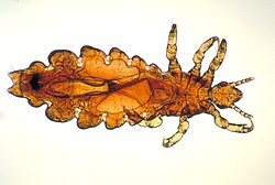
The dorso-ventrally flattened body of the louse is divided into head, thorax and abdomen. On the head, one pair of eyes and one pair of antennae are clearly visible. The mouthparts are adapted to piercing the skin and sucking blood. The legs, with their terminal claws, are adapted to holding the hair-shaft. In males (Fig.1) the front two legs are slightly larger than the other four. This specialized pair of legs is used for holding the female during copulation. Males are slightly smaller than females and are characterized by a pointed end of the abdomen and a well-developed genital apparatus visible inside the abdomen. Females are characterized by two gonopods in the shape of a W at the end of their abdomen (see figure above). Head lice are 1-3 mm in size, varying according to their stage of development. They are usually grayish in color, but can appear reddish-brown soon after a blood-meal.
-
Fig. 1. Male head louse
Egg and Nit Morphology
Louse eggs contain a single embyro, and are attached near the base of a host hair shaft.[1] After hatching, the louse nymph leaves behind its egg shell, still attached to the hair shaft.
The term "nit" most correctly refers to the remnants of a hatched egg.[2] However, "nit" is used variously to refer to a louse egg (regardless of whether it has hatched or not),[1] and/or a non-viable (dead) louse egg.[3]
Biology
During its lifespan of 4 weeks a female louse lays 50-150 eggs (nits). The egg hatches to the first nymphal stage, which after three moltings develop to nymph 2, nymph 3 and eventually to either a male or female louse. Adult lice copulate frequently and the females lay an average of 3-4 eggs daily. A generation lasts for about 1 month. All stages are blood-feeders and they bite the skin 4-5 times daily to feed. During oviposition the female excretes a glue-like substance from a gland located at the posterior end of the body and attaches the eggs on the hair of the host. Although any part of the scalp may be colonized, lice favor the nape of the neck and the area behind the ears, where the eggs are usually laid.
Template:Head louse pediculosis
Treatment
General recommendations for treatment
The number of cases of human louse infestations (or pediculosis) has increased worldwide since the mid-1960s, reaching hundreds of millions annually.[4] There is no product or method which assures 100% destruction of the eggs and hatched lice after a single treatment. However, there are a number of treatment modalities that can be employed with varying degrees of success. These methods include chemical treatments, natural products, combs, shaving, hot air, and silicone-based lotions.
Ancient lice - Use in archaeogenetics
Lice are also important in the field of Archaeogenetics. Because most "modern" human diseases have in fact recently jumped from animals into humans through close agricultural contact, and also given fact that Neolithic human populations were too scattered to support contagious "crowd" diseases, lice (along with such parasites as intestinal tapeworms) are considered to be one of the few ancestral disease infestations of humans and other hominids. As such, analysis of mitochondrial lice DNA has been used to map early human and archaic human migrations and living conditions. Because lice can only survive for a few hours or days without a human host, and because lice species are so specific to certain species or areas of the body, the evolutionary history of lice reveals much about human history. It has been demonstrated, for example, that some varieties of human lice went through a population bottleneck about 100,000 years ago (supporting the Single origin hypothesis), and also that hominid lice lineages diverged around 1.18 million years ago (probably infesting Homo erectus) before re-uniting around 100,000 years ago. This recent merging seems to argue against the Multi-regional origin of modern human evolution and argues instead for a close proximity replacement of archaic humans by a migration of anatomically modern humans, either through inter-breeding, fighting, or being more fit to use available resources.
See also
References
- ^ a b Williams LK, Reichert A, MacKenzie WR, Hightower AW, Blake PA (2001). "Lice, nits, and school policy". Pediatrics. 107 (5): 1011–5. PMID 11331679.
{{cite journal}}: CS1 maint: multiple names: authors list (link) - ^ Burgess IF (2004). "Human lice and their control". Annu. Rev. Entomol. 49: 457–81. doi:10.1146/annurev.ento.49.061802.123253. PMID 14651472.
- ^ Cite error: The named reference
pmid16911370was invoked but never defined (see the help page). - ^ Gratz, N. (1998). "Human lice, their prevalence and resistance to insecticides". Geneva: World Health Organization (WHO).
{{cite journal}}:|access-date=requires|url=(help); Check date values in:|date=(help); Cite journal requires|journal=(help)

