
Dermatomyositis
| Dermatomyositis | |
|---|---|
| Specialty | Rheumatology, immunology, neurology, dermatology |
| Frequency | 0.01022% |
Dermatomyositis (DM) is a connective-tissue disease related to polymyositis (PM) that is characterized by inflammation of the muscles and the skin. While DM most frequently affects the skin and muscles, it is a systemic disorder that may also affect the joints, the esophagus, the lungs, and the heart.[1] In the United States, the incidence of DM is estimated at 5.5 cases per million people.[2]
Signs and symptoms
The main symptoms include skin rash and symmetric proximal muscle weakness and may also be characterized by inflammation of the blood vessels (vasculitis) affecting the skin, muscles, and internal organs.[3]) Individuals affected by dermatomyositis may find it hard to raise their arms to comb their hair or walk up the stairs due to the proximal muscle weakness. Dermatomyositis can be severe enough to affect the muscles needed for speech and/or swallowing and is also known to cause respiratory compromise (due to a variety of mechanisms). Calcinosis can occur in the skin, joints and tissues.
Skin findings occur in dermatomyositis and are generally present at diagnosis. Gottron's sign is an erythematous, scaly eruption occurring in symmetric fashion over the knuckles and interphalangeal joints (can mimic psoriasis). The heliotrope or "lilac" rash[4] is a violaceous eruption on the upper eyelids and in rare cases on the lower eyelids as well, often with itching and swelling (most specific, though uncommon)[clarification needed]. Shawl (or V-) sign is a diffuse, flat, erythematous lesion over the back and shoulders or in a "V" over the posterior neck and back or neck and upper chest, which worsens with UV light. Erythroderma is a flat, red lesion similar to the shawl sign but located in other areas, such as the cheek region and the forehead.
"Mechanic's hands" refers to rough, cracked skin at the tips and lateral aspects of the fingers forming irregular dirty-appearing lines that resemble those seen in a laborer (this is also associated with the anti-synthetase syndrome). See: sclerodactyly. Changes in the scalp that appear similar to psoriasis can also occur. Centripetal flagellate erythema comprises linear, violet-colored streaks on the trunk (possibly caused by itching pruritic skin). Calcinosis cutis (deposition of calcium in the skin) is usually seen in juvenile dermatomyositis, not adult dermatomyositis. Dysphagia (difficulty swallowing) is another feature, occurring in as many as 33% of cases.[citation needed]
-
 Gottron's papules. Erythematous to violaceous raised papules overlying the metacarpal
 -
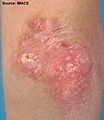 Gottron's papules. Erythematous plaques overlying the elbows in a patient with juvenile dermatomyositis. In some patients, small erythematous plaques may overly the extensor aspects of larger joints, such as the elbows, knees, and medial malleoli. This is considered to be an extended part of the spectrum of Gottron's papules. Note in Figure, a focal area of dystrophic calcification at the site of Gottron's papules, which is indicative of damage, as discussed below.
 -
 Gottron's papules with telangiectasia. Gottron's papules with prominent atrophy, porcelain white scarring, and telangiectasia.
 -
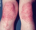 Gottron's sign. Confluent macular erythema with scale confined to the skin overlying the patellae in a girl with juvenile dermatomyositis.
 -
 Gottron's papules showing secondary atrophy and telangiectasia in a girl with severe juvenile dermatomyositis. These changes are seen in association with prominent periungual erythema, a cutaneous finding indicative of ongoing disease activity.
 -
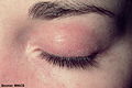 Heliotrope. Confluent macular erythema confined to the upper eyelid, with associated periorbital edema.
 -
 Heliotrope is often associated with periorbital edema and telangiectasias of the upper eyelids. In the resolution stage, atrophy or abnormal pigmentation (hypo- or hyperpigmentation) may be apparent. Heliotrope. Subtle erythema and minimal edema involving both upper eyelids, with extension to the lower eyelids.
 -
 Malar and facial erythema. Striking malar and facial erythema with facial edema and scale represent a recent disease flare in an adult patient with dermatomyositis.
 -
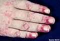
 Linear extensor erythema. Confluent erythema of the skin overlying the interphalangeal and extensor tendons of the hand, with extension to the forearm in a patient with dermatomyositis
 -
 Linear extensor erythema involving the forearm. This image demonstrates linear violaceous discrete and confluent macules, and erosions secondary to excoriation. Pruritus is an under-recognized symptom of active dermatomyositis.


Causes
The cause is unknown, but it may result from an initial viral or bacterial infection (even after treatment or cure of the infection). In cases that follow closely after pregnancy, some research suggests an immune response to lingering fetal cells still circulating in the mother becoming an immune response against the parent, resulting in chronic autoimmune activity. Regardless of the trigger, dermatomyositis may behave as a systemic autoimmune disease. Many people diagnosed with dermatomyositis were previously diagnosed with infectious mononucleosis, Epstein-Barr virus, Chlamydia pneumoniae, psittacosis, and other types of infections. Some cases of dermatomyositis actually "overlap" (i.e. co-exist with or are part of a spectrum that includes) other autoimmune diseases such as Sjögren's syndrome, lupus, scleroderma, or vasculitis. Because of the link between dermatomyositis and autoimmune disease, doctors and patients suspecting dermatomyositis may find it helpful to run an ANA - antinuclear antibody - test, which in cases of a lupus-like nature may be positive (usually from 1:160 to 1:640, with normal ranges at 1:40 or 1:80 and below).[citation needed]
Though several cases of "myositis" were reported as being triggered by the use of various statin drugs (used to control blood cholesterol), the behavior of muscle inflammation linked to statin drugs is different than either dermatomyositis or polymyositis (D.M./P.M.) Muscle biopsies of the statin patients showed rhabdomyolysis, with patterns of degeneration and regeneration of muscle tissue, whereas biopsies of dermatomyositis show "perivascular infiltrates of inflammatory cells,"and muscle regeneration is not present. However, a patient who already has either D.M. or P. M. may need expert advice before taking the risk of statin therapy, since an episode of muscle pain could then be attributed to either the statin or the underlying condition.
High blood levels of creatine kinase (CPK) are often, but not always present in dermatomyositis or polymyositis, causing some patients to be misdiagnosed. The "CPK" levels tend to rise when abnormal inflammation occurs to skeletal muscles and/or other areas of the body. However, since dermatomyositis is highly individualistic, the CPK may be helpful but should not be definitive as a tool for diagnosis. Obviously, extremely high levels on CPK (as seen in rhabdomyolysis) indicate heavy damage to these muscles and other internal organs, such as the kidneys.[clarification needed]. Without treatment, kidney damage occurs and death in the more severe cases. Since dermatomyositis may still damage the heart, intestines, or breathing muscles in a "patchy" way, the definitive test is a muscle biopsy on the affected areas (usually the upper arms/ upper legs). Expertly done E.M.G. testing usually tracks "flagrant" D.M., though again, the neurologist should look for "patches" of involvement in unusual patients who seem to have "normal" tests after having other indications of D.M./P.M.
In his 1988 article, Clinical pathologic correlations of Lyme disease by stage, noted Lyme disease researcher Alan Steere observed, "[...] the perivascular lymphoid infiltrate in clinical myositis does not differ from that seen in polymyositis or dermatomyositis. All of these histologic derangements suggest immunologic damage in response to persistence of the spirochete, however few in number."[5]
Cancer
Between 7 and 30% of dermatomyositis are a paraneoplastic phenomenon, indicating the presence of cancer.[6] The most common associated cancers are ovarian cancer, breast cancer, and lung cancer.[6]
Pathophysiology
The mechanism is conjectured to be complement-mediated damage of microscopic vessels with muscle atrophy and lymphocytic inflammation secondary to tissue ischemia.[7]
Microscopic findings
Cross sections of muscle reveal muscle fascicles with small, shrunken polygonal muscle fibers on the periphery of a fascicle surrounding central muscle fibers of normal, uniform size.[citation needed]
Aggregates of mature lymphocytes with small, dark nuclei and scant cytoplasm are seen surrounding vessels. Other inflammatory cells are distinctly uncommon. Immunohistochemistry can be used to demonstrate that both B- and T-cells are present in approximately equal numbers.[citation needed]
Diagnosis


The diagnosis of dermatomyositis can be confirmed by muscle biopsy, skin biopsy, electromyography (EMG), and blood tests assessing for markers of muscle damage. It should be noted, however, that only muscle biopsy is truly diagnostic; liver enzymes and EMG are relatively non-specific. The two classic microscopic findings seen on muscle biopsy in those affected by dermatomyositis are a mixed B- and T-cell perivascular inflammatory infiltrate and perifascicular muscle fiber atrophy. Spontaneous muscle fibrillation and short polyphasic muscle potentials are characteristic findings on EMG. However, the findings may be variable if the disease affects only patches of muscle and/or overlaps with another autoimmune disease.
Histopathology of skin biopsies from patients with dermatomyositis shows perivascular inflammation, hyperkeratosis in the stratum corneum, epidermal atrophy, follicular plugging, and interface dermatitis (inflammatory cells at or obscuring the dermal-epidermal junction). These changes are not specific for dermatomyositis because they can also be seen in systemic lupus erythematosus.[8]
Dermatomyositis is associated with autoantibodies, especially anti-Mi-2 antibodies, and to a lesser extent anti-Jo-1 antibodies, which are more commonly seen in polymyositis.[citation needed] Other enzymes, specifically creatine phosphokinase (CPK), are the major tool in assessing the progress of the disease and/or the efficacy of treatment.
X-ray findings sometimes include dystrophic calcification in the muscles, and the person may or may not notice small calcium deposits under the skin. Many do not have any calcium deposits of any kind. The rash also may come and go, and may not be dependent on the severity of the muscle involvement at the time.[citation needed]
Classification
Dermatomyositis is considered a muscular dystrophy by the Muscular Dystrophy Association.[9] It is also a type of autoimmune connective tissue disease.[10] It is related to polymyositis and inclusion body myositis.
There is a form of this disorder that strikes children, known as juvenile dermatomyositis (JDM).[citation needed]
Differential diagnosis
Dermatomyositis must be differentiated from other common, lymphocyte predominant inflammatory myopathies. If present, the characteristic perifascicular atrophy makes this distinction trivial.[citation needed]
There is some overlap in the microscopic appearances of different inflammatory myopathies, but some helpful differences are often present.[11] The rimmed vacuoles of inclusion body myositis (IBM) are absent in dermatomyositis. Polymyositis is characterized by diffuse or patchy inflammation of the muscle fascicles, a random pattern of muscle atrophy, and T-cell predominance with T-cells seen invading otherwise viable appearing muscle fibers.[1] Dermatomyositis has been associated with cases of Lyme Disease.[12]
Treatment
This can be treated with steroids and a mild dose of chemotherapy. Specialized exercise therapy may supplement treatment to enhance the quality of life.
Medications to help relieve symptoms include:
- Prednisolone
- Methotrexate
- Mycophenolate
- Hydroxychloroquine
- Intravenous immunoglobulin
- Azathioprine
- Cyclophosphamide
- Tacrolimus
- Infliximab
- Rituximab[13]
Prognosis
Before the advent of modern treatments such as prednisone, intravenous immunoglobulin, plasmapheresis, chemotherapies, and other drugs, the prognosis was poor.[14] Now there are numerous treatments and immunomodulatory drugs. According to published data in 2015 (based on a number of population based cohort studies of adult dermatomyositis and polymyositis patients) the survival rates for dermatomyositis were 70% at 5 years and 55% at 10 years.[15][16] Thus, it is important that treatment begin as soon as possible. The cutaneous manifestations of dermatomyositis may or may not improve with therapy in parallel with the improvement of the myositis. In some patients the weakness and rash resolve together. In others, the two are not linked, with one or the other being more challenging to control. Often, cutaneous disease persists after adequate control of the muscle disease.[8]
Notable cases
- The opera singer Maria Callas (1923 - 1977) suffered from dermatomyositis from 1975 until her death.[17][18]
- The actor Laurence Olivier (1907 – 1989) suffered from dermatomyositis from 1974 until his death.[19]
- The American football running back Ricky Bell (1955 - 1984), the runner-up for the Heisman Trophy in 1976, and the number-one choice in the NFL draft in 1977, died at the age of 29 from heart failure caused by this disease.[20]
- Rob Buckman (1948 - 2011) a doctor, comedian, author, and the president of the Humanist Association of Canada.[21][22]
See also
References
- ^ Dermatomyositis at eMedicine
- ^ http://www.bio.davidson.edu/courses/immunology/students/spring2003/statler/dermatomyositis.htm[full citation needed]
- ^ Bohan, Anthony; Peter, James B.; Bowman, Ralph L.; Pearson, Carl M. (1977). "Computer-assisted analysis of 153 patients with polymyositis and dermatomyositis". Medicine. 56 (4): 255–86. doi:10.1097/00005792-197707000-00001. PMID 327194.
- ^ Mitchell, Richard Sheppard; Kumar, Vinay; Abbas, Abul K.; Fausto, Nelson (2007). Robbins Basic Pathology (8th ed.). Philadelphia: Saunders. p. 151. ISBN 1-4160-2973-7.
- ^ Duray, Paul H.; Steere, Allen C. (1988). "Clinical Pathologic Correlations of Lyme Disease by Stage". Annals of the New York Academy of Sciences. 539: 65–79. doi:10.1111/j.1749-6632.1988.tb31839.x. PMID 2847622.
- ^ a b Di Rollo, D; Abeni, D; Tracanna, M; Capo, A; Amerio, P (October 2014). "Cancer risk in dermatomyositis: a systematic review of the literature". Giornale italiano di dermatologia e venereologia : organo ufficiale, Societa italiana di dermatologia e sifilografia. 149 (5): 525–37. PMID 24975953.
- ^ Benveniste, Olivier; Squier, Waney; Boyer, Olivier; Hilton-Jones, David; Herson, Serge (2004). "Physiopathologie des myopathies inflammatoires primitives". Presse Médicale (in French). 33 (20): 1444–50. doi:10.1016/S0755-4982(04)98952-X. PMID 15611679.
{{cite journal}}: Unknown parameter|trans_title=ignored (|trans-title=suggested) (help) - ^ a b http://www.myositis.org/storage/documents/Publications_for_website/2014_-_Myositis_101_ForDocs_small.pdf[full citation needed]
- ^ "Dermatomyositis".
- ^ "Polymyositis and Dermatomyositis: Autoimmune Disorders of Connective Tissue: Merck Manual Home Edition".
- ^ Nirmalananthan, N; Holton JL; Hanna MG (November 2004). "Is it really myositis? A consideration of the differential diagnosis". Current Opinion in Rheumatology. 16 (6): 684–691. doi:10.1097/01.bor.0000143441.27065.bc. PMID 15577605.
- ^ Dermatomyositis associated with Lyme Disease
- ^ Scheinfeld N (2006). "A review of rituximab in cutaneous medicine". Dermatol. Online J. 12 (1): 3. PMID 16638371.
- ^ Page 285 in: Thomson and Cotton Lecture Notes in Pathology, Blackwell Scientific. Third Edition
- ^ Dobloug, Gerd Cecilie; Garen, Torhild; Brunborg, Cathrine; Gran, Jan Tore; Molberg, Øyvind (2015). "Survival and cancer risk in an unselected and complete Norwegian idiopathic inflammatory myopathy cohort". Seminars in Arthritis and Rheumatism. 45 (3): 301–8. doi:10.1016/j.semarthrit.2015.06.005. PMID 26190563.
- ^ http://arthritis-research.com/content/14/1/R22[full citation needed] Archived 13 December 2014 at the Wayback Machine
- ^ Maria Callas#Fussi and Paolillo report
- ^ "Greek Reporter: 'Maria Callas did not kill herself from grief for Onassis, a rare disease cost her career and life'". GR Reporter. 28 December 2010. Retrieved 1 January 2015.
- ^ "Laurence Olivier Dies: 'The Rest Is Silence'". People Magazine. 24 July 1989. Retrieved 16 July 2012.
- ^ "Forgotten: Ricky Bell". Pro Football Weekly. 8 January 2010. Retrieved 26 January 2010.
- ^ "Rob Buckman obituary". The Guardian Newspaper. 12 October 2011. Retrieved 16 July 2012.
- ^ "Dr. Robert Buckman, renowned oncologist, comedian and Star columnist, dead at 63". The Toronto Star Newspaper. 10 October 2011. Retrieved 16 July 2012.
External links
- The American College of Rheumatology's patient education page on myopathy
- Illustration of Gottron's papules at University of Iowa
- The Johns Hopkins Myositis Center
- Polymyositis and Dermatomyositis, Merck Manual (Professional Version)
- Dourmishev, Lyubomir A.; Dourmishev, Assen Lyubenov (2009). Dermatomyositis: Advances in Recognition, Understanding and Management (2009 ed.). Germany: Springer Verlag. ISBN 978-3642098185.