
File:Development-of-grouped-icEEG-for-the-study-of-cognitive-processing-Video1.ogv
Size of this JPG preview of this OGG file: 800 × 335 pixels. Other resolutions: 320 × 134 pixels | 640 × 268 pixels | 1,799 × 754 pixels.
Original file (Ogg multiplexed audio/video file, Theora/Vorbis, length 30 s, 1,799 × 754 pixels, 1.74 Mbps overall, file size: 6.22 MB)
| This is a file from the Wikimedia Commons. Information from its description page there is shown below. Commons is a freely licensed media file repository. You can help. |
Summary
| Description |
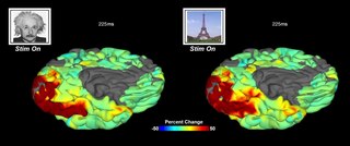
English: Intracranial recordings provide unparalleled insights into rapidly evolving patterns of cortical activity across distributed neural substrates. Electrocorticographic movies of grouped percent change in high-frequency broadband gamma activity (BGA; 60–120 Hz; n = 27 subject) during a face and place-naming task are visualized on the N27 cortical surface model. Hotter colors denote an increase in mid-gamma band power, while cooler colors denote a decrease (color-scale ranges from −50 to 50% change). The movie begins 100 ms before stimulus onset and continues until 700 ms after stimulus onset, in 5 ms steps (stimulus onset at 0 ms). Notably, for face stimuli (left surface) increases in BGA are localized to the lateral aspect of the mid-fusiform sulcus. In contrast, place stimuli produce more widespread activations in the medial aspects of the fusiform gyrus.
Français : Les enregistrements intracrâniens fournissent des aperçus inégalés des modèles d'activité corticale évoluant rapidement dans les réseaux neuraux distribués. L'image électrocorticographique montre ici les changement d'activité exprimés en pourcentage d'activité gamma à large bande à haute fréquence (BGA, 60-120 Hz) à partir de 27 sujets) pour une tâche consistant à reconnaitre un visage ; Les couleurs chaudes montrent une augmentation d'activité dans la bande gamma moyenne, les couleurs froides traduisent une diminution d'activité (l'échelle des couleurs varie de -50 à + 50%). Le film commence 100 ms avant le début du stimulus et se poursuit jusqu'à 700 ms après le début du stimulus, par pas de 5 ms (début du stimulus à 0 ms). Notamment, pour les stimuli du visage (surface gauche), les augmentations de BGA sont localisées à l'aspect latéral du sulcus mi-fusiforme. En revanche, les stimuli de la photo d'un monument (tour eiffel) produisent des activations plus étendues dans les aspects médiaux du gyrus fusiforme |
||
| Date | |||
| Source | Movie 1 from Kadipasaoglu C, Forseth K, Whaley M, Conner C, Rollo M, Baboyan V, Tandon N (2015). "Development of grouped icEEG for the study of cognitive processing". Frontiers in Psychology. DOI:10.3389/fpsyg.2015.01008. PMID 26257673. PMC: 4508923. | ||
| Author | Kadipasaoglu C, Forseth K, Whaley M, Conner C, Rollo M, Baboyan V, Tandon N | ||
| Permission (Reusing this file) |
This file is licensed under the Creative Commons Attribution 4.0 International license.
|
||
| Provenance |
|
File history
Click on a date/time to view the file as it appeared at that time.
| Date/Time | Thumbnail | Dimensions | User | Comment | |
|---|---|---|---|---|---|
| current | 22:22, 12 August 2015 | 30 s, 1,799 × 754 (6.22 MB) | Open Access Media Importer Bot | Automatically uploaded media file from Open Access source. Please report problems or suggestions here. |
File usage
The following pages on the English Wikipedia use this file (pages on other projects are not listed):
Global file usage
The following other wikis use this file:
- Usage on da.wikipedia.org
- Usage on de.wikipedia.org
- Usage on fr.wikipedia.org
- Usage on outreach.wikimedia.org