
File:HLA-A1.png
Size of this preview: 539 × 599 pixels. Other resolutions: 216 × 240 pixels | 548 × 609 pixels.
Original file (548 × 609 pixels, file size: 145 KB, MIME type: image/png)
| This is a file from the Wikimedia Commons. Information from its description page there is shown below. Commons is a freely licensed media file repository. You can help. |
Summary
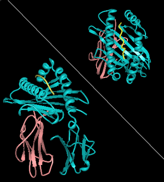
| Description |
English: Rendering of HLA-A1 with MAGE-1 bound peptide. Two views, from the side showing B2 microglobulin (rose) and HLA-A1 (alpha chain, cyan). Top-right view is looking down through the binding site toward the plasma membrane. The image is derived from PDB:1W72 that was presented in the work:Hulsmeyer et al. (2005) A major histocompatibility complex-peptide-restricted antibody and t cell receptor molecules recognize their target by distinct binding modes: crystal structure of human leukocyte antigen (HLA)-A1-MAGE-A1 in complex with FAB-HYB3. J.Biol.Chem. 280: 2972-2980. Image rendered with PDB ProteinWorkshop 1.50. Several aspects of the image removed for clarity reasons. |
| Source | Own work |
| Author | Pdeitiker |
Licensing
| I, the copyright holder of this work, release this work into the public domain. This applies worldwide. In some countries this may not be legally possible; if so: I grant anyone the right to use this work for any purpose, without any conditions, unless such conditions are required by law. |
File history
Click on a date/time to view the file as it appeared at that time.
| Date/Time | Thumbnail | Dimensions | User | Comment | |
|---|---|---|---|---|---|
| current | 20:21, 21 August 2008 | | 548 × 609 (145 KB) | Pdeitiker | {{Information |Description={{en|1=Rendering of HLA-A1 with MAGE-1 bound peptide. Two views, from the side showing B2 microglobulin (rose) and HLA-A1 (alpha chain, cyan). Top-right view is looking down through the binding site toward the plasma membrane. T |
File usage
The following pages on the English Wikipedia use this file (pages on other projects are not listed):
Global file usage
The following other wikis use this file:
- Usage on ar.wikipedia.org
- Usage on pl.wikipedia.org