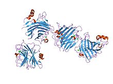
L-type lectin domain
| Lectin_leg-like | |||||||||
|---|---|---|---|---|---|---|---|---|---|
 the crystal structure of the carbohydrate recognition domain of the glycoprotein sorting receptor p58/ergic-53 reveals a novel metal binding site and conformational changes associated with calcium ion binding | |||||||||
| Identifiers | |||||||||
| Symbol | Lectin_leg-like | ||||||||
| Pfam | PF03388 | ||||||||
| Pfam clan | CL0004 | ||||||||
| InterPro | IPR005052 | ||||||||
| SCOP2 | 1gv9 / SCOPe / SUPFAM | ||||||||
| Membranome | 719 | ||||||||
| |||||||||
In molecular biology the L-like lectin domain is a protein domain found in lectins which are similar to the leguminous plant lectins.
Lectins are structurally diverse proteins that bind to specific carbohydrates. This family includes the VIP36 and ERGIC-53 lectins.[1] Although proteins containing this domain were originally identified as a family of animal lectins, there are also yeast representatives.[1]
ERGIC-53 is a 53kDa protein, localised to the intermediate region between the endoplasmic reticulum and the Golgi apparatus (ER-Golgi-Intermediate Compartment, ERGIC). It was identified as a calcium-dependent, mannose-specific lectin.[2] Its dysfunction has been associated with combined factors V and VIII deficiency, suggesting an important and substrate-specific role for ERGIC-53 in the glycoprotein-secreting pathway.[2][3]
The L-like lectin domain has an overall globular shape composed of a beta-sandwich of two major twisted antiparallel beta-sheets. The beta-sandwich comprises a major concave beta-sheet and a minor convex beta-sheet, in a variation of the jelly roll fold.[4][5][6][7]
References[edit]
- ^ a b Fiedler K, Simons K (June 1994). "A putative novel class of animal lectins in the secretory pathway homologous to leguminous lectins". Cell. 77 (5): 625–6. doi:10.1016/0092-8674(94)90047-7. PMID 8205612. S2CID 21111364.
- ^ a b Itin C, Roche AC, Monsigny M, Hauri HP (March 1996). "ERGIC-53 is a functional mannose-selective and calcium-dependent human homologue of leguminous lectins". Mol. Biol. Cell. 7 (3): 483–93. doi:10.1091/mbc.7.3.483. PMC 275899. PMID 8868475.
- ^ Nichols WC, Terry VH, Wheatley MA, Yang A, Zivelin A, Ciavarella N, Stefanile C, Matsushita T, Saito H, de Bosch NB, Ruiz-Saez A, Torres A, Thompson AR, Feinstein DI, White GC, Negrier C, Vinciguerra C, Aktan M, Kaufman RJ, Ginsburg D, Seligsohn U (April 1999). "ERGIC-53 gene structure and mutation analysis in 19 combined factors V and VIII deficiency families". Blood. 93 (7): 2261–6. PMID 10090935.
- ^ Velloso LM, Svensson K, Schneider G, Pettersson RF, Lindqvist Y (May 2002). "Crystal structure of the carbohydrate recognition domain of p58/ERGIC-53, a protein involved in glycoprotein export from the endoplasmic reticulum". J. Biol. Chem. 277 (18): 15979–84. doi:10.1074/jbc.M112098200. PMID 11850423.
- ^ Velloso LM, Svensson K, Pettersson RF, Lindqvist Y (December 2003). "The crystal structure of the carbohydrate-recognition domain of the glycoprotein sorting receptor p58/ERGIC-53 reveals an unpredicted metal-binding site and conformational changes associated with calcium ion binding". J. Mol. Biol. 334 (5): 845–51. doi:10.1016/j.jmb.2003.10.031. PMID 14643651.
- ^ Satoh T, Sato K, Kanoh A, Yamashita K, Yamada Y, Igarashi N, Kato R, Nakano A, Wakatsuki S (April 2006). "Structures of the carbohydrate recognition domain of Ca2+-independent cargo receptors Emp46p and Emp47p". J. Biol. Chem. 281 (15): 10410–9. doi:10.1074/jbc.M512258200. PMID 16439369.
- ^ Satoh T, Cowieson NP, Hakamata W, Ideo H, Fukushima K, Kurihara M, Kato R, Yamashita K, Wakatsuki S (September 2007). "Structural basis for recognition of high mannose type glycoproteins by mammalian transport lectin VIP36" (PDF). J. Biol. Chem. 282 (38): 28246–55. doi:10.1074/jbc.M703064200. PMID 17652092. S2CID 33042130.
