Malate dehydrogenase
| Malate dehydrogenase | |||||||||
|---|---|---|---|---|---|---|---|---|---|
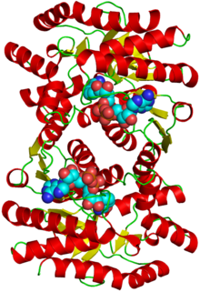
 Structure of the protein with attached cofactors | |||||||||
| Identifiers | |||||||||
| EC no. | 1.1.1.37 | ||||||||
| CAS no. | 9001-64-3 | ||||||||
| Databases | |||||||||
| IntEnz | IntEnz view | ||||||||
| BRENDA | BRENDA entry | ||||||||
| ExPASy | NiceZyme view | ||||||||
| KEGG | KEGG entry | ||||||||
| MetaCyc | metabolic pathway | ||||||||
| PRIAM | profile | ||||||||
| PDB structures | RCSB PDB PDBe PDBsum | ||||||||
| |||||||||
Malate dehydrogenase (EC 1.1.1.37) (MDH) is an enzyme that reversibly catalyzes the oxidation of malate to oxaloacetate using the reduction of NAD+ to NADH. This reaction is part of many metabolic pathways, including the citric acid cycle. Other malate dehydrogenases, which have other EC numbers and catalyze other reactions oxidizing malate, have qualified names like malate dehydrogenase (NADP+).
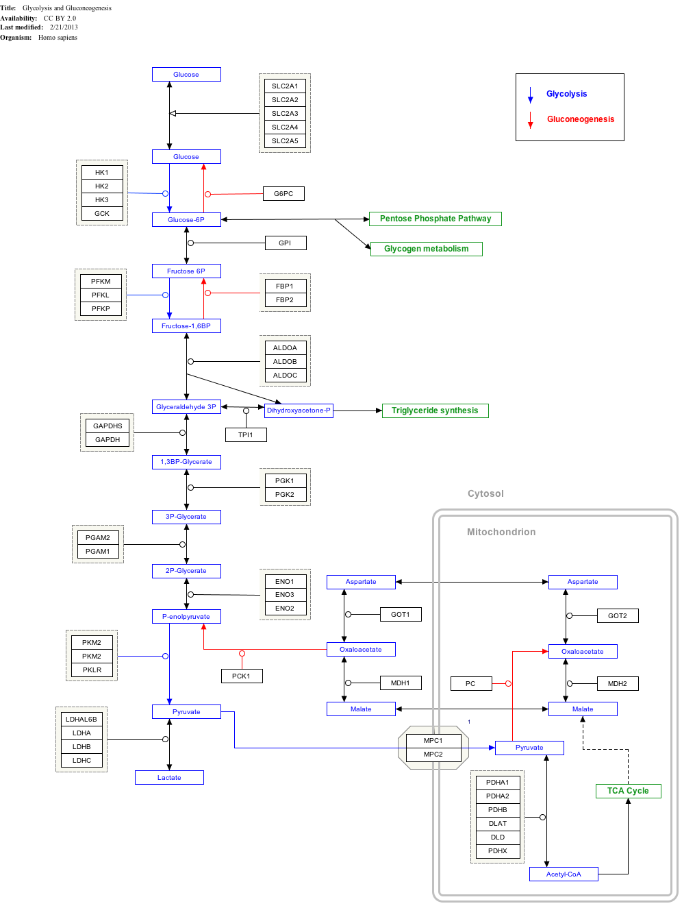
Malate dehydrogenase is also involved in gluconeogenesis, the synthesis of glucose from smaller molecules. Pyruvate in the mitochondria is acted upon by pyruvate carboxylase to form oxaloacetate, a citric acid cycle intermediate. In order to get the oxaloacetate out of the mitochondria, malate dehydrogenase reduces it to malate, and it then traverses the inner mitochondrial membrane. Once in the cytosol, the malate is oxidized back to oxaloacetate by cytosolic malate dehydrogenase. Finally, phosphoenolpyruvate carboxykinase (PEPCK) converts oxaloacetate to phosphoenolpyruvate (PEP).
Isozymes
Several isozymes of malate dehydrogenase exist. There are two main isoforms in eukaryotic cells.[1] One is found in the mitochondrial matrix, participating as a key enzyme in the citric acid cycle that catalyzes the oxidation of malate. The other is found in the cytoplasm, assisting the malate-aspartate shuttle with exchanging reducing equivalents so that malate can pass through the mitochondrial membrane to be transformed into oxaloacetate for further cellular processes.[2]
Humans and most other mammals express the following two malate dehydrogenases:
|
| ||||||||||||||||||||||||||||||||||||||||||||||||||||||||||||
Protein families
|
| ||||||||||||||||||||||||||||||||||||||||||||||||||
Malate dehydrogenases catalyse the interconversion of malate to oxaloacetate. The enzyme participates in the citric acid cycle. The family also contains L-lactate dehydrogenases that catalyse the conversion of L-lactate to pyruvate, the last step in anaerobic glycolysis. L-2-hydroxyisocaproate dehydrogenases are also members of the family. The N-terminus is a Rossmann NAD-binding fold and the C-terminus is an unusual alpha+beta fold.[3][4]
Evolution and structure
In most organisms, malate dehydrogenase exists as a homodimeric molecule and is closely related to lactate dehydrogenase in structure. It is a large protein molecule with subunits weighing between 30 and 35 kDa.[5] Based on the amino acid sequences, it seems that MDH has diverged into two main phylogenetic groups that closely resemble either mitochondrial isozymes or cytoplasmic/chloroplast isozymes.[6] Because the sequence identity of malate dehydrogenase in the mitochondria is more closely related to its prokaryotic ancestors in comparison to the cytoplasmic isozyme, the theory that mitochondria and chloroplasts were developed through endosymbiosis is plausible.[7] It is interesting to note that the amino acid sequences of archaeal MDH are more similar to that of LDH than that of MDH of other organisms. This indicates that there is a possible evolutionary linkage between lactate dehydrogenase and malate dehydrogenase.[8]
Each subunit of the malate dehydrogenase dimer has two distinct domains that vary in structure and functionality. A parallel β-sheet structure makes up the domain, while four β-sheets and one α-helix comprise the central NAD+ binding site. The subunits are held together through extensive hydrogen-bonding and hydrophobic interactions.[9]
Mechanism and activity
The active site of malate dehydrogenase is a hydrophobic cavity within the protein complex that has specific binding sites for the substrate and its coenzyme, NAD+. In its active state, MDH undergoes a conformational change that encloses the substrate to minimize solvent exposure and to position key residues in closer proximity to the substrate.[6] The three residues in particular that comprise a catalytic triad are histidine (His-195), aspartate (Asp-168), both of which work together as a proton transfer system, and arginines (Arg-102, Arg-109, Arg-171), which secure the substrate.[10] Kinetic studies show that MDH enzymatic activity is ordered. NAD+/NADH is bound before the substrate.[11]

Allosteric regulation
Because malate dehydrogenase is closely tied to the citric acid cycle, regulation is highly dependent on TCA products. High malate concentrations stimulate MDH activity, and, in a converse manner, high oxaloacetate concentrations inhibit the enzyme.[12] Citrate can both allosterically activate and inhibit the enzymatic activity of MDH. It inhibits the oxidation of malate when there are low levels of malate and NAD+. However, in the presence of high levels of malate and NAD+, citrate can stimulate the production of oxaloacetate. It is believed that there is an allosteric regulatory site on the enzyme where citrate can bind to and drive the reaction equilibrium in either direction.[13]
Interactive pathway map
Click on genes, proteins and metabolites below to link to respective articles.[§ 1]
- ^ The interactive pathway map can be edited at WikiPathways: "GlycolysisGluconeogenesis_WP534".
References
- ^ Minárik P, Tomásková N, Kollárová M, Antalík M (Sep 2002). "Malate dehydrogenases--structure and function". General Physiology and Biophysics. 21 (3): 257–65. PMID 12537350.
- ^ Musrati RA, Kollárová M, Mernik N, Mikulásová D (Sep 1998). "Malate dehydrogenase: distribution, function and properties". General Physiology and Biophysics. 17 (3): 193–210. PMID 9834842.
- ^ Chapman AD, Cortés A, Dafforn TR, Clarke AR, Brady RL (Jan 1999). "Structural basis of substrate specificity in malate dehydrogenases: crystal structure of a ternary complex of porcine cytoplasmic malate dehydrogenase, alpha-ketomalonate and tetrahydoNAD". Journal of Molecular Biology. 285 (2): 703–12. doi:10.1006/jmbi.1998.2357. PMID 10075524.
- ^ Madern D (Jun 2002). "Molecular evolution within the L-malate and L-lactate dehydrogenase super-family". Journal of Molecular Evolution. 54 (6): 825–40. doi:10.1007/s00239-001-0088-8. PMID 12029364.
- ^ Banaszak, LJ, Bradshaw RA (1975). "Malate dehydrogenase". In Boyer PD (ed.). The Enzymes. Vol. 11 (3rd ed.). New York: Academic Press. pp. 369–396.
{{cite book}}: Cite has empty unknown parameter:|chapterurl=(help)CS1 maint: multiple names: authors list (link) - ^ a b Goward CR, Nicholls DJ (Oct 1994). "Malate dehydrogenase: a model for structure, evolution, and catalysis". Protein Science. 3 (10): 1883–8. doi:10.1002/pro.5560031027. PMC 2142602. PMID 7849603.
- ^ McAlister-Henn L (May 1988). "Evolutionary relationships among the malate dehydrogenases". Trends in Biochemical Sciences. 13 (5): 178–81. doi:10.1016/0968-0004(88)90146-6. PMID 3076279.
- ^ Cendrin F, Chroboczek J, Zaccai G, Eisenberg H, Mevarech M (Apr 1993). "Cloning, sequencing, and expression in Escherichia coli of the gene coding for malate dehydrogenase of the extremely halophilic archaebacterium Haloarcula marismortui". Biochemistry. 32 (16): 4308–13. doi:10.1021/bi00067a020. PMID 8476859.
- ^ Hall MD, Levitt DG, Banaszak LJ (Aug 1992). "Crystal structure of Escherichia coli malate dehydrogenase. A complex of the apoenzyme and citrate at 1.87 A resolution". Journal of Molecular Biology. 226 (3): 867–82. doi:10.1016/0022-2836(92)90637-Y. PMID 1507230.
- ^ Lamzin VS, Dauter Z, Wilson KS (May 1994). "Dehydrogenation through the looking-glass". Nature Structural Biology. 1 (5): 281–2. doi:10.1038/nsb0594-281. PMID 7664032.
- ^ Shows TB, Chapman VM, Ruddle FH (Dec 1970). "Mitochondrial malate dehydrogenase and malic enzyme: Mendelian inherited electrophoretic variants in the mouse". Biochemical Genetics. 4 (6): 707–18. doi:10.1007/BF00486384. PMID 5496232.
- ^ Mullinax TR, Mock JN, McEvily AJ, Harrison JH (Nov 1982). "Regulation of mitochondrial malate dehydrogenase. Evidence for an allosteric citrate-binding site". The Journal of Biological Chemistry. 257 (22): 13233–9. PMID 7142142.
- ^ Gelpí JL, Dordal A, Montserrat J, Mazo A, Cortés A (Apr 1992). "Kinetic studies of the regulation of mitochondrial malate dehydrogenase by citrate". The Biochemical Journal. 283 (Pt 1): 289–97. doi:10.1042/bj2830289. PMC 1131027. PMID 1567375.
Further reading
- Guha A, Englard S, Listowsky I (Feb 1968). "Beef heart malic dehydrogenases. VII. Reactivity of sulfhydryl groups and conformation of the supernatant enzyme". The Journal of Biological Chemistry. 243 (3): 609–15. PMID 5637713.
{{cite journal}}: Cite has empty unknown parameter:|author-name-separator=(help) - McReynolds MS, Kitto GB (Feb 1970). "Purification and properties of Drosophila malate dehydrogenases". Biochimica et Biophysica Acta. 198 (2): 165–75. doi:10.1016/0005-2744(70)90048-3. PMID 4313528.
{{cite journal}}: Cite has empty unknown parameter:|author-name-separator=(help) - Wolfe RG, Neilands JB (Jul 1956). "Some molecular and kinetic properties of heart malic dehydrogenase". The Journal of Biological Chemistry. 221 (1): 61–9. PMID 13345798.
{{cite journal}}: Cite has empty unknown parameter:|author-name-separator=(help)
External links
- Malate+dehydrogenase at the U.S. National Library of Medicine Medical Subject Headings (MeSH)

