
Ovarian artery
| Ovarian artery | |
|---|---|
 Arteries of the female reproductive tract: uterine artery, ovarian artery and vaginal arteries. | |
 Ovary of a sheep. 1. ovary 2. tertiary follicle 3. proper ovarial ligament 4. fallopian tube 5. A. and V. ovarica | |
| Details | |
| Source | abdominal aorta |
| Branches | tubal branches of ovarian artery |
| Vein | ovarian vein |
| Supplies | ovaries, uterus |
| Identifiers | |
| Latin | arteria ovarica |
| TA98 | A12.2.12.086F |
| TA2 | 4285 |
| FMA | 14761 |
| Anatomical terminology | |
In human anatomy, the ovarian artery is a blood vessel that supplies oxygenated blood to the ovary. It arises from the abdominal aorta below the renal artery, and does not pass out of the abdominal cavity. It can be found in the suspensory ligament of the ovary, anterior to the ovarian vein and ureter.[1]
Relationship to internal spermatic
It only exists in females. The ovarian arteries are the corresponding arteries in the female to the testicular artery in the male. They are shorter than the internal spermatics.
The origin and course of the first part of each artery are the same as those of the internal spermatic, but on arriving at the upper opening of the lesser pelvis the ovarian artery passes inward, between the two layers of the ovariopelvic ligament and of the broad ligament of the uterus, to be distributed to the ovary.
Branches
Small branches are given to the ureter and the uterine tube, and one passes on to the side of the uterus, and unites with the uterine artery.
Other offsets are continued on the round ligament of the uterus, through the inguinal canal, to the integument of the labium majus and groin.
It commonly anastomoses (connects with) the uterine artery.[2]
Additional images
-
 The abdominal aorta and its branches.
 -
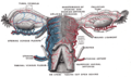 Vessels of the uterus and its appendages, rear view.
 -
 Uterus and right broad ligament, seen from behind.


References
- ^ II, Anne M.R. Agur, Arthur F. Dalley (2009). Grant's atlas of anatomy. Philadelphia: Wolters Kluwer Health/Lippincott Williams & Wilkins. p. 223. ISBN 978-0-7817-9604-0.
{{cite book}}: CS1 maint: multiple names: authors list (link) - ^ Lampmann LE, Smeets AJ, Lohle PN. Uterine fibroids: targeted embolization, an update on technique. Abdom Imaging. 2003 Oct 31; PMID 14583818.
External links
- "Anastomoses Between Utero - Ovarian Arteries, Variations" at anatomyatlases.org
- pelvis at The Anatomy Lesson by Wesley Norman (Georgetown University) (uterus)
- figures/chapter_35/35-9.HTM: Basic Human Anatomy at Dartmouth Medical School
This cardiovascular system article is a stub. You can help Wikipedia by expanding it. |