
Sclera
| Sclera | |
|---|---|
 Schematic diagram of the human eye. | |
| Details | |
| Artery | anterior ciliary arteries, long posterior ciliary arteries, short posterior ciliary arteries |
| Identifiers | |
| MeSH | D012590 |
| TA98 | A15.2.02.002 |
| TA2 | 6750 |
| FMA | 58269 |
| Anatomical terminology | |
The sclera, also known as the white or white of the eye, is the opaque (usually white, though certain animals, such as horses and lizards, can have black sclera), fibrous, protective, outer layer of the eye containing collagen and elastic fiber.[1] In the development of the embryo, the sclera is derived from the neural crest.[2] In children, it is thinner and shows some of the underlying pigment, appearing slightly blue. In the elderly, fatty deposits on the sclera can make it appear slightly yellow.
Human eyes are somewhat unique in the animal kingdom in that the sclera is very plainly visible at all times (except when the eye is closed). This is not just due to the white color of the human sclera, which many other species share, but also to the fact that the human iris is relatively small and comprises a significantly smaller portion of the exposed eye surface compared to other animals. It is theorized that this adaptation evolved because of our social nature as the eye became a useful communication tool in addition to a sensory organ. It's believed that the conspicuous sclera of the human eye makes it easier for one individual to infer where another individual is looking, increasing the efficacy of this particular form of nonverbal communication. Animal researchers have also found that, in the course of their domestication, dogs have also developed the ability to pick up visual cues from the eyes of humans, making them one of only two species known to seek visual cues from another individual's eyes. Interestingly enough, dogs do not seem to use this form of communication with one another and only look for visual information from the eyes of humans.[3]
Structure
The sclera forms the posterior five-sixths of the connective tissue coat of the globe. It is continuous with the dura mater and the cornea, and maintains the shape of the globe, offering resistance to internal and external forces, and provides an attachment for the extraocular muscle insertions. The sclera is perforated by many nerves and vessels passing through the posterior scleral foramen, the hole that is formed by the optic nerve. At the optic disc the outer two-thirds of the sclera continues with the dura mater (outer coat of the brain) via the dural sheath of the optic nerve. The inner third joins with some choroidal tissue to form a plate (lamina cribrosa) across the optic nerve with perforations through which the optic fibers (fasciculi) pass. The thickness of the sclera varies from 1mm at the posterior pole to 0.3 mm just behind the rectus muscle insertions. The sclera's blood vessels are mainly on the surface. Along with the vessels of the conjunctiva (which is a thin layer covering the sclera), those of the sclera render the inflamed eye bright red.[4]
In many vertebrates, the sclera is reinforced with plates of cartilage or bone, together forming a circular structure called the scleral ring. In primitive fish, this ring consists of four plates, but the number is lower in many living ray-finned fishes, and much higher in lobe-finned fishes, various reptiles, and birds. The ring has disappeared in many groups, including living amphibians, some reptiles and fish, and all mammals.[5]
The eyes of all non-human primates are dark with small, barely visible sclera. See Cooperative eye hypothesis
Histology
The collagen of the sclera is continuous with the cornea. From outer to innermost, the four layers of the sclera are:
The sclera is opaque due to the irregularity of the collagen fibers, as opposed to the near-uniform thickness and parallel arrangement of the corneal collagen. Moreover, the cornea bears more mucopolysaccharide (a carbohydrate that has among its repeating units a nitrogenous sugar, hexosamine) to embed the fibrils.
The cornea, unlike the sclera, has 5 layers. The middle, thickest layer is also called the stroma. The sclera, like the cornea, contains a basal endothelium, above which there is the lamina fusca, containing a high count of pigment cells. [4]
Sometimes, very small gray-blue spots can appear on the sclera, a harmless condition called scleral melanocytosis.
Additional images

-
 Interior of anterior half of bulb of eye.
 -
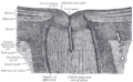 The terminal portion of the optic nerve and its entrance into the eyeball, in horizontal section.
 -
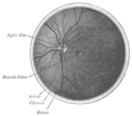 The interior of the posterior half of the left eyeball.


References
- ^ Cassin, B. and Solomon, S. Dictionary of Eye Terminology. Gainesville, Florida: Triad Publishing Company, 1990.
- ^ Hermann D. Schubert. Anatomy of the Orbit http://www.nyee.edu/pdf/schubert.pdf
- ^ Director and Producer: Dan Child, Executive Producer: Andrew Kohen (2010-01-06). "The Secret Life of the Dog". Horizon. BBC. BBC2.
{{cite episode}}: Unknown parameter|serieslink=ignored (|series-link=suggested) (help) - ^ a b "eye, human."Encyclopædia Britannica from Encyclopædia Britannica 2006 Ultimate Reference Suite DVD 2009
- ^ Romer, Alfred Sherwood; Parsons, Thomas S. (1977). The Vertebrate Body. Philadelphia, PA: Holt-Saunders International. p. 461. ISBN 0-03-910284-X.
External links
- Histology image: 08008loa – Histology Learning System at Boston University
- Atlas image: eye_1 at the University of Michigan Health System - "Sagittal Section Through the Eyeball"
| Fibrous tunic (outer) |
|  | |||||
|---|---|---|---|---|---|---|---|
| Uvea / vascular tunic (middle) |
| ||||||
| Retina (inner) |
| ||||||
| Anatomical regions of the eye |
| ||||||
| Other | |||||||
| Illusions |
| |
|---|---|---|
| Popular culture |
| |
| Related | ||