User:Cruithne9/sandbox
Birds[edit]


1. skull
2. cervical vertebrae
3. furcula
4. coracoid
5. vertebral ribs
6. keel
7. patella
8. tarsometatarsus
9. digits
10. tibia (tibiotarsus)
11. fibia (tibiotarsus)
12. femur
13. ischium (innominate)
14. pubis (innominate)
15. illium (innominate)
16. caudal vertebrae
17. pygostyle
18. synsacrum
19. scapula
20. dorsal vertebrae
21. humerus
22. ulna
23. radius
24. carpus (carpometacarpus)
25. metacarpus (carpometacarpus)
26. digits
27. alula
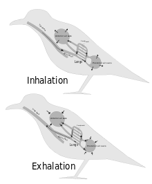
The respiratory system of birds differs significantly from that found in mammals. Firstly they have rigid lungs which do not expand and contract during the breathing cycle. Instead an extensive system of air sacs (Fig. 15) distributed throughout their bodies act as the bellows drawing environmental air into the sacs, and expelling the spent air after it has passed through the lungs (Fig. 16).[1] Birds also do not have diaphragms or pleural cavities.
Inhalation and exhalation are brought about by alternately increasing and decreasing the the volume of the entire thoraco-abdominal cavity (or coelom) using both their abdominal and costal muscles.[2][3][4] During inhalation the muscles attached to the vertebral ribs contract angling them forwards and outwards. This pushes the sternal ribs, to which they are attached at almost right angles, downwards and forwards, taking the sternum, or keel, in the same direction. This increases both the vertical and transverse diameters of thoracic portion of the trunk. The forward and downward movement of particularly the posterior end of the sternum pulls the abdominal wall downwards, increasing the volume of that region of the trunk as well.[2] The increase in volume of the entire trunk cavity reduces the air pressure in all the thoraco-abdominal air sacs, causing them to fill with air as will be described below.
During exhalation the external oblique muscle which is attached to the sternum and vertebral ribs anteriorly, and to the pelvis posteriorly (forming part of the abdominal wall) reverses the inhalatory movement, while compressing the abdominal contents, thus increasing the pressure in all the air sacs. Air is therefore expelled from the respiratory system in the act of exhalation.[2]

During inhalation air enters the trachea via the nostrils and mouth, and continues to just beyond the syrinx at which point the trachea branches into two primary bronchi, going to the two lungs. The primary bronchi enter the lungs to become the intrapulmonary bronchi, which give off a set of parallel branches called ventrobronchi and, a little further on, an equivalent set of dorsobronchi.[2] The ends of the intrapulmonary bronchi discharge air into the posterior air sacs at the caudal end of the bird. Each pair of dorso-ventrobronchi is connected by a large number of parallel microscopic air capillaries (or parabronchi) where gas exchange occurs.[2] As the bird inhales, tracheal air flows through the intrapulmonary bronchi into the posterior air sacs, as well as into the dorsobronchi, but not into the ventrobronchi (Fig. 16). This is due to the bronchial architecture which directs the inhaled air away from the openings of the ventrobronchi, into the continuation of the intrapulmonary bronchus towards the dorsobronchi and posterior air sacs[6][7][8]). From the dorsobronchi the inhaled air flows through the parabronchi (and therefore the gas exchanger) to the ventrobronchi from where the air can only escape into the expanding anterior air sacs. So, during inhalation, both the posterior and anterior air sacs expand,[2] the posterior air sacs filling with fresh inhaled air, while the anterior air sacs fill with "spent" (oxygen-poor) air that has just passed through the lungs.
During exhalation the pressure in the posterior air sacs (which were filled with fresh air during inhalation) increases due to the contraction of the oblique muscle described above. The aerodynamics of the interconnecting openings from the posterior air sacs to the the dorsobronchi and intrapulmonary bronchi ensures that the air leaves these sacs in the direction of the lungs (via the dorsobronchi), rather than returning down the intrapulmonary bronchi (Fig. 16).[6][8] From the the dorsobronchi the fresh air from the posterior air sacs flows through the parabronchi (in the same direction as occurred during inhalation) into ventrobronchi. The air passages connecting the ventrobronchi and anterior air sacs to the intrapulmonary bronchi direct the "spent", oxygen poor air from these two organs to the trachea from where it escapes to the exterior.[2] Oxygenated air therefore flows constantly (during the entire breathing cycle) in a single direction through the parabronchi.[9]
The blood flow through the bird lung is at right angles to the flow of air through the parabronchi, forming a cross-current flow exchange system (Fig. 17).[2][5][1] The partial pressure of oxygen in the parabronchi declines along their lengths as O2 diffuses into the blood. The blood capillaries leaving the exchanger near the entrance of airflow take up more O2 than do the capillaries leaving near the exit end of the parabronchi. When the contents of all capillaries mix, the final partial pressure of oxygen of the mixed pulmonary venous blood is higher than that of the exhaled air,[2][5] but is nevertheless less than half that of the inhaled air,[2] thus achieving roughly the same systemic arterial blood partial pressure of oxygen as mammals do with their bellows-type lungs.[2]
- ^ a b Campbell, Neil A. (1990). Biology (2nd ed. ed.). Redwood City, Calif.: Benjamin/Cummings Pub. Co. pp. 836–844. ISBN 0-8053-1800-3.
{{cite book}}:|edition=has extra text (help) - ^ a b c d e f g h i j k l Ritchson, G. "BIO 554/754 – Ornithology: Avian respiration". Department of Biological Sciences, Eastern Kentucky University. Retrieved 2009-04-23.
- ^ Storer, Tracy I.; Usinger, R. L.; Stebbins, Robert C.; Nybakken, James W. (1997). General Zoology (sixth ed.). New York: McGraw-Hill. pp. 752–753. ISBN 0-07-061780-5.
- ^ Romer, Alfred Sherwood (1970). The Vertebrate body (Fourth ed.). Philadelphia: W.B. Saunders. pp. 323–324. ISBN 0-7216-7667-7.
- ^ a b c Scott, Graham R. (2011). "Commentary: Elevated performance: the unique physiology of birds that fly at high altitudes". Journal of Experimental Biology. 214: 2455–2462. doi:10.1242/jeb.052548.
- ^ a b Maina, John N. (2005). The lung air sac system of birds development, structure, and function ; with 6 tables. Berlin: Springer. pp. 3.2–3.3 "Lung", "Airway (Bronchiol) System" 66–82. ISBN 978-3-540-25595-6.
- ^ Krautwald-Junghanns, Maria-Elisabeth; et al. (2010). Diagnostic Imaging of Exotic Pets: Birds, Small Mammals, Reptiles. Germany: Manson Publishing. ISBN 978-3-89993-049-8.
- ^ a b Sturkie, P.D. (1976). Avian Physiology. New York: Springer Verlag. p. 201. doi:10.1007/978-1-4612-4862-0. ISBN 978-1-4612-9335-4.
- ^ Ritchison, Gary. "Ornithology (Bio 554/754):Bird Respiratory System". Eastern Kentucky University. Retrieved 2007-06-27.

