
Sphenopalatine foramen
| Sphenopalatine foramen | |
|---|---|
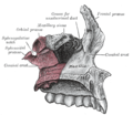
 Medial wall of left orbit. (Sphenopalatine foramen labeled in upper right.) | |
 Left palatine bone. Posterior aspect. Enlarged. (Sphenopalatine foramen labeled in upper right.) | |
| Details | |
| Identifiers | |
| Latin | foramen sphenopalatinum |
| TA98 | A02.1.00.097 |
| TA2 | 502 |
| FMA | 53144 |
| Anatomical terms of bone | |
The sphenopalatine foramen is a fissure of the skull that connects the nasal cavity and the pterygopalatine fossa. It gives passage to the sphenopalatine artery, nasopalatine nerve, and the superior nasal nerve (all passing from the pterygopalatine fossa into the nasal cavity).[1]
Structure[edit]
The processes of the superior border of the palatine bone are separated by the sphenopalatine notch, which is converted into the sphenopalatine foramen by the under surface of the body of the sphenoid.[citation needed]
The sphenopalatine foramen is situated posterior to the middle nasal meatus orbital process of palatine bone, anterior to the sphenoidal process of palatine bone, inferior to the body and concha[clarification needed] of the sphenoid bone, and superior to the superior margin of the perpendicular plate of palatine bone.[1]
Relations[edit]
The ethmoid crest (a reliable surgical landmark) is situated anterior to the sphenopalatine foramen.[1]
Additional images[edit]
-
 Articulation of left palatine bone with maxilla.
 -
 Left palatine bone. Nasal aspect. Enlarged.


References[edit]
- ^ a b c Standring, Susan (2020). Gray's Anatomy: The Anatomical Basis of Clinical Practice (42th ed.). New York. p. 690. ISBN 978-0-7020-7707-4. OCLC 1201341621.
{{cite book}}: CS1 maint: location missing publisher (link)
Sources[edit]
![]() This article incorporates text in the public domain from page 168 of the 20th edition of Gray's Anatomy (1918)
This article incorporates text in the public domain from page 168 of the 20th edition of Gray's Anatomy (1918)
External links[edit]
- lesson9 at The Anatomy Lesson by Wesley Norman (Georgetown University) (nasalwallbones) (#10)
- Anatomy photo:22:os-1108 at the SUNY Downstate Medical Center
This human musculoskeletal system article is a stub. You can help Wikipedia by expanding it. |