
Protist locomotion: Difference between revisions
Epipelagic (talk | contribs) See also + video |
Epipelagic (talk | contribs) Swimming speeds |
||
| Line 1: | Line 1: | ||
[[Protist]]s are the [[eukaryote]]s that cannot be classified as plants, fungi or animals. They are mostly [[unicellular]] and [[microscopic]]. Many unicellular protists, particularly [[protozoan]]s, are [[motile]] and can [[Aquatic locomotion#Micro-organisms|generate movement]] using [[flagella]], [[cilia]] or [[pseudopod]]s. Cells which use flagella for movement are usually referred to as [[flagellate]]s, cells which use cilia are usually referred to as [[ciliate]]s, and cells which use pseudopods are usually referred to as [[amoeba]] or [[amoeboid]]s. Other protists are [[Sessility (motility)|not motile]], and consequently have no built-in movement mechanism. |
[[Protist]]s are the [[eukaryote]]s that cannot be classified as plants, fungi or animals. They are mostly [[unicellular]] and [[microscopic]]. Many unicellular protists, particularly [[protozoan]]s, are [[motile]] and can [[Aquatic locomotion#Micro-organisms|generate movement]] using [[flagella]], [[cilia]] or [[pseudopod]]s. Cells which use flagella for movement are usually referred to as [[flagellate]]s, cells which use cilia are usually referred to as [[ciliate]]s, and cells which use pseudopods are usually referred to as [[amoeba]] or [[amoeboid]]s. Other protists are [[Sessility (motility)|not motile]], and consequently have no built-in movement mechanism. |
||
== |
==Modes of locomotion== |
||
{|class="wikitable" |
{|class="wikitable" |
||
! colspan=9 | {{centre|Protists according to how they move}} |
! colspan=9 | {{centre|Protists according to how they move}} |
||
| Line 79: | Line 79: | ||
==Amoebas== |
==Amoebas== |
||
[[File:Collection Penard MHNG Specimen 05bis-1-1 Amoeba proteus.tif|thumb|Amoeba with ingested diatoms]] |
[[File:Collection Penard MHNG Specimen 05bis-1-1 Amoeba proteus.tif|thumb|Amoeba with ingested diatoms]] |
||
{{clear}} |
|||
==Swimming speeds== |
|||
[[File:Cladogram for some unicellular eukaryotes.webp.jpg|thumb|upright=1.8| {{center|'''Cladogram for some unicellular eukaryote examples'''}} Aquatic organisms (living in marine, brackish, or freshwater environments) have their branches drawn in blue while parasitic organisms have their branches drawn in red. Ciliates are indicated by an asterisk after their names. For each phylum marked in bold font, a representative organism has been sketched next to its name.<ref>{{cite journal |doi = 10.1073/pnas.1423041112}}</ref><ref name=Lisicki2019 />]] |
|||
Unicellular eukaryotes comprise a vast, diverse group of organisms that covers virtually all environments and habitats, displaying a menagerie of shapes and forms. Hundreds of species of the ciliate genus ''[[Paramecium]]''{{hsp}}<ref>{{Cite book|url=https://books.google.com/books?id=JB_pBwAAQBAJ&pg=PA1|title = The Biology of Paramecium|isbn = 9781475703726|last1 = Wichterman|first1 = R.|date = 6 December 2012}}</ref> or flagellated ''[[Euglena]]''{{hsp}}<ref>{{cite book |doi = 10.1002/9780470015902.a0001964.pub3|chapter = Euglena|title = eLS|year = 2011|last1 = Buetow|first1 = Dennis E.|isbn = 978-0470016176}}</ref> are found in marine, brackish, and freshwater reservoirs; the green algae ''[[Chlamydomonas]]'' is distributed in soil and fresh water world-wide;<ref>{{Cite book|url=https://books.google.com/books?id=xTjJGV5GWY0C&pg=PP1|title = The Chlamydomonas Sourcebook: Introduction to Chlamydomonas and Its Laboratory Use: Volume 1|isbn = 9780080919553|last1 = Harris|first1 = Elizabeth H.|date = 7 March 2009}}</ref> parasites from the genus ''[[Giardia]]'' colonize intestines of several vertebrates.<ref>{{cite journal |doi = 10.1128/CMR.14.3.447-475.2001|title = Biology of Giardia lamblia|year = 2001|last1 = Adam|first1 = Rodney D.|journal = Clinical Microbiology Reviews|volume = 14|issue = 3|pages = 447–475|pmid = 11432808|pmc = 88984}}</ref> One of the shared features of these organisms is their motility, crucial for nutrient acquisition and avoidance of danger.<ref>{{Cite book|url=https://books.google.com/books?id=1HBTDwAAQBAJ&pg=PP1|title = Cell Movements: From Molecules to Motility|isbn = 9781136844355|last1 = Bray|first1 = Dennis|date = 2 November 2000}}</ref> In the process of evolution, single-celled organisms have developed in a variety of directions, and thus their rich morphology results in a large spectrum of swimming modes.<ref>{{cite book |doi = 10.1007/978-94-009-5812-8_4|chapter = The movement of eukaryotic cells|title = Motility of Living Cells|year = 1980|last1 = Cappuccinelli|first1 = P.|pages = 59–74|isbn = 978-0-412-15770-7}}</ref><ref name=Lisicki2019>{{cite journal |doi = 10.7554/eLife.44907|title = Swimming eukaryotic microorganisms exhibit a universal speed distribution|year = 2019|last1 = Lisicki|first1 = Maciej|last2 = Velho Rodrigues|first2 = Marcos F.|last3 = Goldstein|first3 = Raymond E.|last4 = Lauga|first4 = Eric|journal = eLife|volume = 8|pmid = 31310238|pmc = 6634970|arxiv = 1907.00906}} [[File:CC-BY icon.svg|50px]] Material was copied from this source, which is available under a [https://creativecommons.org/licenses/by/4.0/ Creative Commons Attribution 4.0 International License].</ref> |
|||
Many swimming eukaryotes actuate tail-like appendages called flagella or cilia in order to generate the required thrust (Sleigh, 1975). This is achieved by actively generating deformations along the flagellum, giving rise to a complex waveform. The flagellar axoneme itself is a bundle of nine pairs of microtubule doublets surrounding two central microtubules, termed the '9 + 2' structure (Nicastro et al., 2005), and cross-linking dynein motors, powered by ATP hydrolysis, perform mechanical work by promoting the relative sliding of filaments, resulting in bending deformations.<ref name=Lisicki2019 /> |
|||
Although eukaryotic flagella exhibit a diversity of forms and functions (Moran et al., 2014), two large families, ‘flagellates’ and ‘ciliates’, can be distinguished by the shape and beating pattern of their flagella. Flagellates typically have a small number of long flagella distributed along the bodies, and they actuate them to generate thrust. The set of observed movement sequences includes planar undulatory waves and traveling helical waves, either from the base to the tip, or in the opposite direction (Jahn and Votta, 1972; Brennen and Winet, 1977). Flagella attached to the same body might follow different beating patterns, leading to a complex locomotion strategy that often relies also on the resistance the cell body poses to the fluid. In contrast, propulsion of ciliates derives from the motion of a layer of densely-packed and collectively-moving cilia, which are short hair-like flagella covering their bodies. The seminal review paper of Brennen and Winet (1977) lists a few examples from both groups, highlighting their shape, beat form, geometric characteristics and swimming properties. Cilia may also be used for transport of the surrounding fluid, and their cooperativity can lead to directed flow generation. In higher organisms this can be crucial for internal transport processes, as in cytoplasmic streaming within plant cells (Allen and Allen, 1978), or the transport of ova from the ovary to the uterus in female mammals (Lyons et al., 2006).<ref name=Lisicki2019 /> |
|||
[[File:Swimming speeds of flagellates and ciliates.jpg|thumb|upright=1.8|left| {{center|'''Swimming speeds of flagellates and ciliates{{hsp}}<ref name=Lisicki2019 />'''<br/><small>Linear distribution of swimming speed data</small>}}]] |
|||
[[File:Normalized curves for flagellate and ciliate swimming speeds.jpg|thumb|upright=1.8| {{center|Fitted log-normal curves for flagellate and ciliate swimming speeds}} Each of these speeds have been normalized by multiplying then by their distribution median. The normalized distributions show remarkable similarity and uncertainty of estimation.<ref name=Lisicki2019 />]] |
|||
{{clear}} |
{{clear}} |
||
Revision as of 06:11, 8 June 2021
Protists are the eukaryotes that cannot be classified as plants, fungi or animals. They are mostly unicellular and microscopic. Many unicellular protists, particularly protozoans, are motile and can generate movement using flagella, cilia or pseudopods. Cells which use flagella for movement are usually referred to as flagellates, cells which use cilia are usually referred to as ciliates, and cells which use pseudopods are usually referred to as amoeba or amoeboids. Other protists are not motile, and consequently have no built-in movement mechanism.
Modes of locomotion
Protists according to how they move
| ||||||||
|---|---|---|---|---|---|---|---|---|
| Type of protist | Movement mechanism | Description | Example | Other examples | ||||
| Motile | Flagellates | 
|
A flagellum (Latin for whip) is a lash-like appendage that protrudes from the cell body of some protists (as well as some bacteria). Flagellates use from one to several flagella for locomotion and sometimes as feeding and sensory organelle. | 
|
Cryptophytes | All dinoflagellates and nanoflagellates (choanoflagellates, silicoflagellates, most green algae)[1][2] (Other protists go through a phase as gametes when they have temporary flagellum – some radiolarians, foraminiferans and Apicomplexa)
| ||
| Ciliates | 
|
A cilium (Latin for eyelash) is a tiny flagellum. Ciliates use multiple cilia, which can number in many hundreds, to power themselves through the water. | .jpg)
|
Paramecium bursaria click to see cilia |
Foraminiferans, and some marine amoebae, ciliates and flagellates. | |||
| Amoebas (amoeboids) |

|
Pseudopods (Greek for false feet) are lobe-like appendages which amoebas use to anchor to a solid surface and pull themselves forward. They can change their shape by extending and retracting these pseudopods.[3] |  |
Amoeba | Found in every major protist lineage. Amoeboid cells occur among the protozoans, but also in the algae and the fungi.[4][5] | |||
| Not motile | none
|
 |
Diatom | Diatoms, coccolithophores, and non‐motile species of Phaeocystis[2] Among protozoans the parasitic Apicomplexa are non‐motile. | ||||
Flagellates

Flagella are used in prokaryotes (archaea and bacteria) as well as protists. In addition, both flagella and cilia are widely used in eukaryotic cells (plant and animal) apart from protists.
The regular beat patterns of eukaryotic cilia and flagella generates motion on a cellular level. Examples range from the propulsion of single cells such as the swimming of spermatozoa to the transport of fluid along a stationary layer of cells such as in a respiratory tract. Though eukaryotic flagella and motile cilia are ultrastructurally identical, the beating pattern of the two organelles can be different. In the case of flagella, the motion is often planar and wave-like, whereas the motile cilia often perform a more complicated three-dimensional motion with a power and recovery stroke.
Eukaryotic flagella—those of animal, plant, and protist cells—are complex cellular projections that lash back and forth. Eukaryotic flagella are classed along with eukaryotic motile cilia as undulipodia[6] to emphasize their distinctive wavy appendage role in cellular function or motility. Primary cilia are immotile, and are not undulipodia.

Cryptaulax, Abollifer, Bodo, Rhynchomonas, Kittoksia, Allas, and Metromonas [7]
-
 Green algal flagellate (Chlamydomonas)
.jpg)
Ciliates

Ciliates generally have hundreds to thousands of cilia that are densely packed together in arrays. Like the flagella, the cilia are powered by specialised molecular motors. An efficient forward stroke is made with a stiffened flagellum, followed by an inefficient backward stroke made with a relaxed flagellum. During movement, an individual cilium deforms as it uses the high-friction power strokes and the low-friction recovery strokes. Since there are multiple cilia packed together on an individual organism, they display collective behaviour in a metachronal rhythm. This means the deformation of one cilium is in phase with the deformation of its neighbor, causing deformation waves that propagate along the surface of the organism. These propagating waves of cilia are what allow the organism to use the cilia in a coordinated manner to move. A typical example of a ciliated microorganism is the Paramecium, a one-celled, ciliated protozoan covered by thousands of cilia. The cilia beating together allow the Paramecium to propel through the water at speeds of 500 micrometers per second.[8]
-
 Paramecium feeding on bacteria
 -
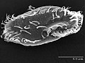 The ciliate Oxytricha trifallax with cilia clearly visible


| External videos | |
|---|---|
Amoebas

Swimming speeds

Unicellular eukaryotes comprise a vast, diverse group of organisms that covers virtually all environments and habitats, displaying a menagerie of shapes and forms. Hundreds of species of the ciliate genus Paramecium [11] or flagellated Euglena [12] are found in marine, brackish, and freshwater reservoirs; the green algae Chlamydomonas is distributed in soil and fresh water world-wide;[13] parasites from the genus Giardia colonize intestines of several vertebrates.[14] One of the shared features of these organisms is their motility, crucial for nutrient acquisition and avoidance of danger.[15] In the process of evolution, single-celled organisms have developed in a variety of directions, and thus their rich morphology results in a large spectrum of swimming modes.[16][10]
Many swimming eukaryotes actuate tail-like appendages called flagella or cilia in order to generate the required thrust (Sleigh, 1975). This is achieved by actively generating deformations along the flagellum, giving rise to a complex waveform. The flagellar axoneme itself is a bundle of nine pairs of microtubule doublets surrounding two central microtubules, termed the '9 + 2' structure (Nicastro et al., 2005), and cross-linking dynein motors, powered by ATP hydrolysis, perform mechanical work by promoting the relative sliding of filaments, resulting in bending deformations.[10]
Although eukaryotic flagella exhibit a diversity of forms and functions (Moran et al., 2014), two large families, ‘flagellates’ and ‘ciliates’, can be distinguished by the shape and beating pattern of their flagella. Flagellates typically have a small number of long flagella distributed along the bodies, and they actuate them to generate thrust. The set of observed movement sequences includes planar undulatory waves and traveling helical waves, either from the base to the tip, or in the opposite direction (Jahn and Votta, 1972; Brennen and Winet, 1977). Flagella attached to the same body might follow different beating patterns, leading to a complex locomotion strategy that often relies also on the resistance the cell body poses to the fluid. In contrast, propulsion of ciliates derives from the motion of a layer of densely-packed and collectively-moving cilia, which are short hair-like flagella covering their bodies. The seminal review paper of Brennen and Winet (1977) lists a few examples from both groups, highlighting their shape, beat form, geometric characteristics and swimming properties. Cilia may also be used for transport of the surrounding fluid, and their cooperativity can lead to directed flow generation. In higher organisms this can be crucial for internal transport processes, as in cytoplasmic streaming within plant cells (Allen and Allen, 1978), or the transport of ova from the ovary to the uterus in female mammals (Lyons et al., 2006).[10]

Linear distribution of swimming speed data

See also
References
- ^ Dawson, Scott C; Paredez, Alexander R (2013). "Alternative cytoskeletal landscapes: cytoskeletal novelty and evolution in basal excavate protists". Current Opinion in Cell Biology. 25 (1): 134–141. doi:10.1016/j.ceb.2012.11.005. PMC 4927265. PMID 23312067.
- ^ a b Atkinson, A.; Polimene, L.; Fileman, E.S.; Widdicombe, C.E.; McEvoy, A.J.; Smyth, T.J.; Djeghri, N.; Sailley, S.F.; Cornwell, L.E. (2018). ""Comment. What drives plankton seasonality in a stratifying shelf sea? Some competing and complementary theories"]" (PDF). Limnology and Oceanography. 63 (6): 2877–2884. Bibcode:2018LimOc..63.2877A. doi:10.1002/lno.11036.
- ^ Singleton, Paul (2006). Dictionary of Microbiology and Molecular Biology, 3rd Edition, revised. Chichester, UK: John Wiley & Sons. pp. 32. ISBN 978-0-470-03545-0.
- ^ David J. Patterson. "Amoebae: Protists Which Move and Feed Using Pseudopodia". Tree of Life web project.
- ^ "The Amoebae". The University of Edinburgh. Archived from the original on 10 June 2009.
- ^ A Dictionary of Biology, 2004, accessed 2011-01-01.
- ^ Patterson, David J. (2000) "Flagellates: Heterotrophic Protists With Flagella" Tree of Life.
- ^ Lauga, Eric; Thomas R Powers (25 August 2009). "The hydrodynamics of swimming microorganisms". Reports on Progress in Physics. 72 (9): 096601. arXiv:0812.2887. Bibcode:2009RPPh...72i6601L. doi:10.1088/0034-4885/72/9/096601. S2CID 3932471.
- ^ . doi:10.1073/pnas.1423041112.
{{cite journal}}: Cite journal requires|journal=(help); Missing or empty|title=(help) - ^ a b c d e f Lisicki, Maciej; Velho Rodrigues, Marcos F.; Goldstein, Raymond E.; Lauga, Eric (2019). "Swimming eukaryotic microorganisms exhibit a universal speed distribution". eLife. 8. arXiv:1907.00906. doi:10.7554/eLife.44907. PMC 6634970. PMID 31310238.
{{cite journal}}: CS1 maint: unflagged free DOI (link) Material was copied from this source, which is available under a Creative Commons Attribution 4.0 International License.
- ^ Wichterman, R. (6 December 2012). The Biology of Paramecium. ISBN 9781475703726.
- ^ Buetow, Dennis E. (2011). "Euglena". eLS. doi:10.1002/9780470015902.a0001964.pub3. ISBN 978-0470016176.
- ^ Harris, Elizabeth H. (7 March 2009). The Chlamydomonas Sourcebook: Introduction to Chlamydomonas and Its Laboratory Use: Volume 1. ISBN 9780080919553.
- ^ Adam, Rodney D. (2001). "Biology of Giardia lamblia". Clinical Microbiology Reviews. 14 (3): 447–475. doi:10.1128/CMR.14.3.447-475.2001. PMC 88984. PMID 11432808.
- ^ Bray, Dennis (2 November 2000). Cell Movements: From Molecules to Motility. ISBN 9781136844355.
- ^ Cappuccinelli, P. (1980). "The movement of eukaryotic cells". Motility of Living Cells. pp. 59–74. doi:10.1007/978-94-009-5812-8_4. ISBN 978-0-412-15770-7.