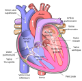
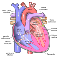
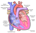
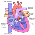
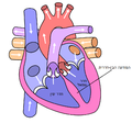
File:Diagram of the human heart (cropped) pt.svg
Appearance

Size of this PNG preview of this SVG file: 577 × 599 pixels. Other resolutions: 231 × 240 pixels | 462 × 480 pixels | 739 × 768 pixels | 986 × 1,024 pixels | 1,971 × 2,048 pixels | 667 × 693 pixels.
Original file (SVG file, nominally 667 × 693 pixels, file size: 280 KB)
File history
Click on a date/time to view the file as it appeared at that time.
| Date/Time | Thumbnail | Dimensions | User | Comment | |
|---|---|---|---|---|---|
| current | 12:22, 16 July 2021 |  | 667 × 693 (280 KB) | Jmarchn | Validation W3C |
| 21:54, 14 August 2018 |  | 667 × 693 (299 KB) | Jmarchn | Add pericardium. Add papillary muscles and chordae tendinae. Add cardiac skeleton. Inferior vena cava more wide. Add aorta in bottom. Add source veins of superior vena cava. Brachiocephalic trunk more wide and separated. Added shadows. Left main pulmonary artery with its first division. | |
| 04:37, 25 January 2007 |  | 670 × 670 (38 KB) | PbBR8498 | {{Information |Description=Diagrama do coração humano (cortado) |Source=Diagram_of_the_human_heart_(cropped).svg |Date=01/25/2007 |Author= |Permission= |other_versions= <gallery> Image:Diagram of the human heart (cropped).svg|English version Image:Diagr |
File usage
No pages on the English Wikipedia use this file (pages on other projects are not listed).
Global file usage
The following other wikis use this file:
- Usage on es.wikipedia.org
- Usage on gl.wikipedia.org
- Usage on pt.wikipedia.org
- Corpo humano
- Ventrículo
- Aurícula
- Nó sinusal
- Veia cava superior
- Veia cava inferior
- Válvula tricúspide
- Válvula pulmonar
- Válvula mitral
- Válvula aórtica
- Artéria pulmonar
- Fisiologia humana
- Predefinição:Info/Artéria
- Veia cava
- Síndrome da veia cava superior
- Predefinição:Info/Artéria/doc
- Usuário:Ernesto Capiquila
- Usuário:EduardoFP7/Testes/1
- Insuficiência pulmonar


