
Lacrimal gland
| Lacrimal gland | |
|---|---|
 Lacrimal apparatus of the right eye. The lacrimal gland is to the upper left. The right side of the picture is towards the nose. | |
 Tear system. a = lacrimal gland b = superior lacrimal punctum c = superior lacrimal canal d = lacrimal sac e = inferior lacrimal punctum f = inferior lacrimal canal g = nasolacrimal canal | |
| Details | |
| Artery | lacrimal artery |
| Nerve | lacrimal nerve, Zygomatic nerve via Communicating branch |
| Identifiers | |
| Latin | glandula lacrimalis |
| TA98 | A15.2.07.057 |
| TA2 | 6846 |
| FMA | 59101 |
| Anatomical terminology | |
The lacrimal glands are paired almond-shaped glands, one for each eye, that secrete the aqueous layer of the tear film. They are situated in the upper, outer portion of each orbit, in the lacrimal fossa of the orbit formed by the frontal bone.[1] Inflammation of the lacrimal glands is called dacryoadenitis. The lacriminal gland produces tears which then flow into canals that lead to the lacriminal sac. From this sac, the tears drain through a passage into the nose. [citation needed]
Anatomists divide the gland into two sections. The smaller palpebral portion, lies close to the eye, along the inner surface of the eyelid; if the upper eyelid is everted, the palpebral portion can be seen.
The orbital portion contains fine interlobular ducts that unite to form 3 - 5 main excretory ducts, joining 5 - 7 ducts in the palpebral portion before the secreted fluid may enter on the surface of the eye. Tears secreted collect in the fornix conjunctiva of the upper lid, and pass over the eye surface to the lacrimal puncta, small holes found at the inner corner of the eyelids. These pass the tears on to the lacrimal sac, in turn to the nasolacrimal duct, which dumps them out into the nose.[2]
Microanatomy
The lacrimal gland is a compound tubuloacinar gland, it is made up of many lobules separated by connective tissue, each lobule contains many acini. The acini contain only serous cells and produce a watery serous secretion.
Each acinus consists of a grape-like mass of lacrimal gland cells with their apices pointed to a central lumen.
The central lumen of many of the units converge to form intralobular ducts, and then unite to from interlobular ducts. The gland lacks striated ducts.
Innervation
The parasympathetic nerve supply originates from the lacrimal nucleus of the facial nerve in the pons. Just distal to the geniculate ganglion, the facial nerve gives off the greater petrosal nerve. This nerve carries the parasympathetic secretomotor fibers through the pterygoid canal, where it joins the deep petrosal nerve (containing postganglionic sympathetic fibers from the superior cervical ganglion) to form the nerve of the pterygoid canal (vidian nerve). This nerve travels through the pterygoid canal to the pterygopalatine ganglion. Here the fibers synapse and postganglionic fibers join the fibers of the Maxillary Nerve, which travels through the inferior orbital fissure. Once it has traversed this opening, the parasympathetic secretomotor fibers branch off with the zygomatic nerve and then branch off again, joining with the lacrimal branch of the ophthalmic division of CN V, which supplies sensory innervation to the lacrimal gland along with the eyelid and conjunctiva.
The sympathetic postganglionic fibres originate from the superior cervical ganglion. They travel as a periarteriolar plexus with the middle meningeal artery, before they merge and form the deep petrosal nerve, which joins the greater petrosal nerve in the pterygoid canal. Together, greater petrosal and deep petrosal nerves form the nerve of the pterygoid canal (vidian nerve) and reach the pterygopalatine ganglion in the pterygopalatine fossa. In contrast to their parasympathetic counterparts, sympathetic fibers do not synapse in the pterygopalatine ganglion, having done so already in the sympathetic trunk. However, they continue to course with the parasympathetic fibers innervating the lacrimal gland.
Blood supply
The lacrimal artery, derived from the ophthalmic artery supplies the lacrimal gland. Venous blood returns via the superior ophthalmic vein.
Nerve supply
The lacrimal nerve, derived from the ophthalmic nerve supplies the lacrimal gland
Applied anatomy
- The upper part of the lacrimal sac is covered by the medial palpabral ligament. Hence the abscesses within the sac bulge below the medial palpabrel ligament, where it should be incised for letting out the pus.
- The angular vein is in front of the sac and it should be take care during incising the sac. Between the lacrimal sac and the fascia coverning the sac there is the collection of venous plexus present hence the incising causes considerable bleeding.
- Around the nasolacrimal duct there is the rich plexus of veins, in the form of an erectile tissue, which may engorge and cause obstruction to the duct.
Pathology
Additional images
-
 The ophthalmic artery and its branches.
 -
 Nerves of the orbit. Seen from above.
 -
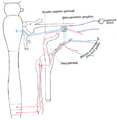 Sympathetic connections of the sphenopalatine and superior cervical ganglia.
 -
 The tarsal glands, etc., seen from the inner surface of the eyelids.
 -
 Alveoli of lacrimal gland.


See also
References
- ^ Clinically Oriented Anatomy, Moore, Dalley & Agur.
- ^ "eye, human."Encyclopædia Britannica. 2010. Encyclopædia Britannica 2010 Ultimate Reference Suite DVD 2010
External links
- Template:EMedicineDictionary
- lesson3 at The Anatomy Lesson by Wesley Norman (Georgetown University) (orbit2)