
Visceral pleura
| Pulmonary pleura | |
|---|---|
 A transverse section of the thorax, showing the contents of the middle and the posterior mediastinum. The pleural and pericardial cavities are exaggerated since normally there is no space between parietal and visceral pleura and between pericardium and heart. | |
| Details | |
| Nerve | pulmonary plexus |
| Identifiers | |
| Latin | pleura visceralis, pleura pulmonalis |
| TA98 | A07.1.02.002 |
| TA2 | 3325 |
| TH | H3.05.03.0.00008 |
| FMA | 9734 |
| Anatomical terminology | |
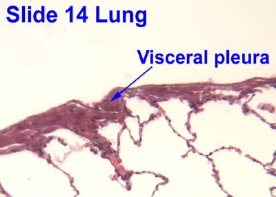
Each lung is invested by an exceedingly delicate serous membrane, the pleura, which is arranged in the form of a closed invaginated sac. A portion of the serous membrane covers the surface of the lung and dips into the fissures between its lobes; it is called the pulmonary pleura (or visceral pleura). The visceral pleura is derived from mesoderm.
The visceral pleura is attached directly to the lungs, as opposed to the parietal pleura, which is attached to the opposing thoracic cavity. The space between these two delicate membranes is known as the intrapleural space (pleural cavity). Contraction of the diaphragm causes a negative pressure within this space and forces the lungs to expand, resulting in passive exhalation and active inhalation. This process can be made forceful through the contraction of the external intercostal muscles, forcing the rib cage to expand and aiding to the negative pressure within the intrapleural space, which causes the lungs to fill with air.
Additional Images
-
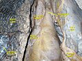 Visceral and parietal pleura.Deep dissection. Anterior view.

External links
- . GPnotebook https://www.gpnotebook.co.uk/simplepage.cfm?ID=74121277.
{{cite web}}: Missing or empty|title=(help) - thoraxlesson2 at The Anatomy Lesson by Wesley Norman (Georgetown University)
- Atlas image: lung_lymph at the University of Michigan Health System - "Transverse section through lung"
- Histology image: 14_15 at the University of Oklahoma Health Sciences Center - "Lung"
- MedEd at Loyola Grossanatomy/thorax0/thor_lec/thor6.html
![]() This article incorporates text in the public domain from page 1087 of the 20th edition of Gray's Anatomy (1918)
This article incorporates text in the public domain from page 1087 of the 20th edition of Gray's Anatomy (1918)
This respiratory system article is a stub. You can help Wikipedia by expanding it. |