LE cell: Difference between revisions
No edit summary |
No edit summary |
||
| Line 1: | Line 1: | ||
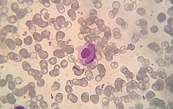
[[File:LECell.jpg|thumb|250px|Microphotograph of a macrophage that has phagocytized a lymphocyte (LE cell). May Grünwald Giemsa stain.]] |
[[File:LECell.jpg|thumb|250px|Microphotograph of a macrophage that has phagocytized a lymphocyte (LE cell). May Grünwald Giemsa stain.]] |
||
An '''LE cell''' is a [[neutrophil]] or [[macrophage]] that has phagocytized (engulfed) the denatured nuclear material of another cell.<ref>http://www.merriam-webster.com/medical/le%20cell</ref> The denatured material is an absorbed [[hematoxylin body]] (also called an LE body).<ref>[http://www.similima.com/pathology/path11.html similima.com > Autoimmunity] By Muhammed Muneer. Retrieved Mars 2011</ref> |
An '''LE cell''' ('''L'''upus '''E'''rythromatosus cell) is a [[neutrophil]] or [[macrophage]] that has phagocytized (engulfed) the denatured nuclear material of another cell.<ref>http://www.merriam-webster.com/medical/le%20cell</ref> The denatured material is an absorbed [[hematoxylin body]] (also called an LE body).<ref>[http://www.similima.com/pathology/path11.html similima.com > Autoimmunity] By Muhammed Muneer. Retrieved Mars 2011</ref> |
||
They are a characteristic of [[lupus erythematosus]],<ref>http://www.drugsfreelist.com/health_articles/4002/LE_Cell_Test__Lupus_erythematosus_test__</ref> but also found in similar connective tissue disorders. |
They are a characteristic of [[lupus erythematosus]],<ref>http://www.drugsfreelist.com/health_articles/4002/LE_Cell_Test__Lupus_erythematosus_test__</ref> but also found in similar connective tissue disorders or some [[autoimmune diseases]] like in severe [[rheumatoid arthritis]]. LE cells could be drug-induced, following treatment with [[methyldopa]].<ref>{{Cite book|url=https://books.google.de/books?id=PHPx140zdowC&pg=PA295|title=District Laboratory Practice in Tropical Countries|last=Cheesbrough|first=Monica|date=2000-10-26|publisher=Cambridge University Press|isbn=9780521665452|language=en}}</ref> |
||
The LE cell was discovered in [[bone marrow]] in 1948 by Malcolm McCallum Hargraves (1903–1982), a Physician and Practicing Histologist at the [[Mayo Clinic]] ''et al.''<ref>Hargraves M, Richmond H, Morton R. ''Presentation of two bone marrow components, the tart cell and the LE cell.'' Mayo Clin Proc 1948;27:25–28.</ref> |
The LE cell was discovered in [[bone marrow]] in 1948 by Malcolm McCallum Hargraves (1903–1982), a Physician and Practicing Histologist at the [[Mayo Clinic]] ''et al.''<ref>Hargraves M, Richmond H, Morton R. ''Presentation of two bone marrow components, the tart cell and the LE cell.'' Mayo Clin Proc 1948;27:25–28.</ref> |
||
Classically, the LE cell is analyzed microscopically, but it is also possible to investigate this phenomenon by [[flow cytometry]].<ref name="pmid15115310">{{cite journal|last=Böhm|first=Ingrid|title=Flow Cytometric Analysis of the LE Cell Phenomenon|journal=Autoimmunity|date=1 January 2004|volume=37|issue=1|pages=37–44|doi=10.1080/08916930310001630325|pmid=15115310}}</ref> |
Classically, the LE cell is analyzed microscopically, but it is also possible to investigate this phenomenon by [[flow cytometry]].<ref name="pmid15115310">{{cite journal|last=Böhm|first=Ingrid|title=Flow Cytometric Analysis of the LE Cell Phenomenon|journal=Autoimmunity|date=1 January 2004|volume=37|issue=1|pages=37–44|doi=10.1080/08916930310001630325|pmid=15115310}}</ref> |
||
LE cells shouldn't be confused with [[Tart cell]]s which have engulfed nuclear material, but with a visible chromatin rather than homogenous appearance. <ref>{{Cite book|url=https://link.springer.com/chapter/10.1007/978-1-4939-1477-7_2|title=Diagnostic Cytopathology Board Review and Self-Assessment|last=Li|first=Qing Kay|last2=Khalbuss|first2=Walid E.|date=2015|publisher=Springer, New York, NY|isbn=9781493914760|pages=179|language=en|doi=10.1007/978-1-4939-1477-7_2}}</ref> |
|||
==References== |
==References== |
||
Revision as of 00:24, 11 October 2017

An LE cell (Lupus Erythromatosus cell) is a neutrophil or macrophage that has phagocytized (engulfed) the denatured nuclear material of another cell.[1] The denatured material is an absorbed hematoxylin body (also called an LE body).[2]
They are a characteristic of lupus erythematosus,[3] but also found in similar connective tissue disorders or some autoimmune diseases like in severe rheumatoid arthritis. LE cells could be drug-induced, following treatment with methyldopa.[4]
The LE cell was discovered in bone marrow in 1948 by Malcolm McCallum Hargraves (1903–1982), a Physician and Practicing Histologist at the Mayo Clinic et al.[5]
Classically, the LE cell is analyzed microscopically, but it is also possible to investigate this phenomenon by flow cytometry.[6]
LE cells shouldn't be confused with Tart cells which have engulfed nuclear material, but with a visible chromatin rather than homogenous appearance. [7]
References
- ^ http://www.merriam-webster.com/medical/le%20cell
- ^ similima.com > Autoimmunity By Muhammed Muneer. Retrieved Mars 2011
- ^ http://www.drugsfreelist.com/health_articles/4002/LE_Cell_Test__Lupus_erythematosus_test__
- ^ Cheesbrough, Monica (2000-10-26). District Laboratory Practice in Tropical Countries. Cambridge University Press. ISBN 9780521665452.
- ^ Hargraves M, Richmond H, Morton R. Presentation of two bone marrow components, the tart cell and the LE cell. Mayo Clin Proc 1948;27:25–28.
- ^ Böhm, Ingrid (1 January 2004). "Flow Cytometric Analysis of the LE Cell Phenomenon". Autoimmunity. 37 (1): 37–44. doi:10.1080/08916930310001630325. PMID 15115310.
- ^ Li, Qing Kay; Khalbuss, Walid E. (2015). Diagnostic Cytopathology Board Review and Self-Assessment. Springer, New York, NY. p. 179. doi:10.1007/978-1-4939-1477-7_2. ISBN 9781493914760.
