
Nasal bone
| Nasal bone | |
|---|---|
 Nasal bone visible at center, in dark green. | |
 Cartilages of the nose. Side view. (Nasal bone visible at upper left.) | |
| Details | |
| Identifiers | |
| Latin | os nasale |
| MeSH | D009295 |
| TA98 | A02.1.10.001 |
| TA2 | 748 |
| FMA | 52745 |
| Anatomical terms of bone | |
The nasal bones are two small oblong bones, varying in size and form in different individuals; they are placed side by side at the middle and upper part of the face, and form, by their junction, "the bridge" of the nose.
Each has two surfaces and four borders.
Surfaces
The outer surface is concavoconvex from above downward, convex from side to side; it is covered by the Procerus and Compressor naris, and perforated about its center by a foramen, for the transmission of a small vein.
The inner surface is concave from side to side, and is traversed from above downward, by a groove for the passage of a branch of the nasociliary nerve.
Articulations
The nasal articulates with four bones: two of the cranium, the frontal and ethmoid, and two of the face, the opposite nasal and the maxilla.
Additional images
-
 Orbital bones
 -
 Lateral wall of nasal cavity, showing ethmoid bone in position.
 -
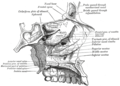
 Articulation of nasal and lacrimal bones with maxilla.
 -
 Right nasal bone. Outer surface.
 -
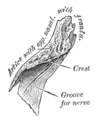 Right nasal bone. Inner surface.
 -
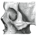 Sphenoid bone visible center right.
 -
 Side view of the skull.
 -
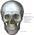 The skull from the front.
 -
 Horizontal section of nasal and orbital cavities.
 -
 Medial wall of left orbit.
 -
 Sagittal section of skull.
 -
 Roof, floor, and lateral wall of left nasal cavity.


See also
External links
- Template:EMedicineDictionary
- Anatomy figure: 22:02-07 at Human Anatomy Online, SUNY Downstate Medical Center - "Anterior view of skull."
- Anatomy photo:29:st-0206 at the SUNY Downstate Medical Center - "Orbits and Eye: Bones"
- Anatomy figure: 33:01-03 at Human Anatomy Online, SUNY Downstate Medical Center - "The bones of the lateral nasal wall."
- Template:RocheLexicon
![]() This article incorporates text in the public domain from page 156 of the 20th edition of Gray's Anatomy (1918)
This article incorporates text in the public domain from page 156 of the 20th edition of Gray's Anatomy (1918)