Neocortex
| Neocortex | |
|---|---|
| Identifiers | |
| MeSH | D019579 |
| NeuroNames | 757 |
| NeuroLex ID | birnlex_2547 |
| TA98 | A14.1.09.304 A14.1.09.307 |
| TA2 | 5532 |
| FMA | 62429 |
| Anatomical terms of neuroanatomy | |

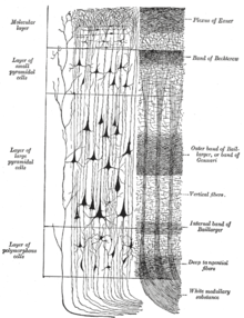
The neocortex (Latin for "new bark" or "new rind"), also called the neopallium ("new mantle") and isocortex ("equal rind"), is a part of the brain of mammals. It is the outer layer of the cerebral hemispheres, and made up of six layers, labelled I to VI (with VI being the innermost and I being the outermost). The neocortex is part of the cerebral cortex (along with the archicortex and paleocortex, which are cortical parts of the limbic system). In humans, it is involved in higher functions such as sensory perception, generation of motor commands, spatial reasoning, conscious thought and language.[1]
Anatomy
The neocortex consists of the grey matter, or neuronal cell bodies and unmyelinated fibers, surrounding the deeper white matter (myelinated axons) in the cerebrum.
The neocortex is smooth in rodents and other small mammals, whereas in primates and other larger mammals it has deep grooves (sulci) and wrinkles (gyri). These folds allow the surface area of the neocortex to increase far beyond what could otherwise be fit in the same size skull. All human brains have the same overall pattern of main gyri and sulci, although they differ in detail from one person to another. The mechanism by which the gyri form during embryogenesis is not entirely clear, and there are several competing hypotheses that explain gyrification. Axonal tension,[2] cortical buckling,[3] or differences in cellular proliferation rates in different areas of the cortex[4] during embryonic development may contribute to the formation of gyri.
The neocortex contains two primary types of neurons, excitatory pyramidal neurons (~80% of neocortical neurons) and inhibitory interneurons (~20%). The structure of the neocortex is relatively uniform (hence the alternative names "iso-" and "homotypic" cortex), consisting of six horizontal layers segregated principally by cell type and neuronal connections. However, there are many exceptions to this uniformity; for example, the motor cortex lacks layer IV. There is some canonical circuitry within the cortex; for example, pyramidal neurons in the upper layers II and III project their axons to other areas of neocortex, while those in the deeper layers V and VI project primarily out of the cortex, e.g. to the thalamus, brainstem, and spinal cord. Neurons in layer IV receive all of the synaptic connections from outside the cortex (mostly from thalamus), and themselves make short-range, local connections to other cortical layers. Thus, layer IV receives all incoming sensory information and distributes it to the other layers for further processing.
The neurons of the neocortex are also arranged in vertical structures called neocortical columns. These are patches of the neocortex with a diameter of about 0.5 mm (and a depth of 2 mm). Each column typically responds to a sensory stimulus representing a certain body part or region of sound or vision. These columns are similar, and can be thought of as the basic repeating functional units of the neocortex. In humans, the neocortex consists of about a half-million of these columns, each of which contains approximately 70,000 neurons.[5]
The neocortex is derived embryonically from the dorsal telencephalon, which is the rostral part of the forebrain. The neocortex is divided into frontal, parietal, occipital, and temporal lobes, which perform different functions. For example, the occipital lobe contains the primary visual cortex, and the temporal lobe contains the primary auditory cortex. Further subdivisions or areas of neocortex are responsible for more specific cognitive processes. In humans, the frontal lobe contains areas devoted to abilities that are enhanced in or unique to our species, such as complex language processing localized to the ventrolateral prefrontal cortex (Broca's area). In humans and other primates, social and emotional processing is localized to the orbitofrontal cortex. (See Cerebral cortex and Cerebrum.)
Evolution
The neocortex is the newest part of the cerebral cortex to evolve (hence the prefix "neo"); the other parts of the cerebral cortex are the paleocortex and archicortex, collectively known as the allocortex. The cellular organization of the allocortex is different from the six-layer structure mentioned above. In humans, 90% of the cerebral cortex is neocortex[citation needed].
The six-layer cortex appears to be a distinguishing feature of mammals; it has been found in the brains of all mammals, but not in any other animals.[1] There is some debate,[6][7] however, as to the cross-species nomenclature for neocortex. In avians, for instance, there are clear examples of cognitive processes that are thought to be neocortical in nature, despite the lack of the distinctive six-layer neocortical structure.[8] In a similar manner, reptiles, such as turtles, have primary sensory cortices. A consistent, alternative name has yet to be agreed upon.
Neocortex ratio
The neocortex ratio of a species is the ratio of the size of the neocortex to the rest of the brain. A high neocortex ratio is thought to correlate with a number of social variables such as group size and the complexity of social mating behaviors.[9] (See Dunbar's number) Humans have a large neocortex as a percentage of total brain matter when compared with other mammals. For example, there is only a 30:1 ratio of neocortical gray matter to the size of the medulla in the brainstem of chimpanzees, while the ratio is 60:1 in humans.[10]
See also
- List of regions in the human brain
- Blue Brain, a project to produce a computer simulation of a neocortical column and eventually a whole neocortex
- Memory-prediction framework, a theory of the neocortex function by Jeff Hawkins and related software models
- Model of the neocortex by the Brain Engineering Laboratory at Dartmouth College
- Comparative Neuroscience at Wikiversity
References
- ^ a b Attention: This template ({{cite pmid}}) is deprecated. To cite the publication identified by PMID 21729779, please use {{cite journal}} with
|pmid=21729779instead. - ^ Van Essen, DC (Jan 23, 1997). "A tension-based theory of morphogenesis and compact wiring in the central nervous system" (PDF). Nature. 385 (6614): 313–8. doi:10.1038/385313a0. PMID 9002514.
- ^ Richman, David (4 July 1975). "Mechanical model of brain convolutional development". Science. 189 (4196): 18–21. doi:10.1126/science.1135626. PMID 1135626.
- ^ Ronan, L. (29 March 2013). "Differential Tangential Expansion as a Mechanism for Cortical Gyrification". Cereb. Cortex. doi:10.1093/cercor/bht082. PMID 23542881.
{{cite journal}}: Unknown parameter|coauthors=ignored (|author=suggested) (help) - ^ bluebrain.epfl.ch
- ^ Jarvis, Erich D.; Güntürkün, Onur; Bruce, Laura; Csillag, András; Karten, Harvey; Kuenzel, Wayne; Medina, Loreta; Paxinos, George; et al. (2005). "Opinion: Avian brains and a new understanding of vertebrate brain evolution". Nature Reviews Neuroscience. 6 (2): 151–9. doi:10.1038/nrn1606. PMC 2507884. PMID 15685220.
- ^ Reiner, Anton; Perkel, David J.; Bruce, Laura L.; Butler, Ann B.; Csillag, András; Kuenzel, Wayne; Medina, Loreta; Paxinos, George; et al. (2004). "Revised nomenclature for avian telencephalon and some related brainstem nuclei". The Journal of Comparative Neurology. 473 (3): 377–414. doi:10.1002/cne.20118. PMC 2518311. PMID 15116397.
- ^ Prior, Helmut; Schwarz, Ariane; Güntürkün, Onur (2008). De Waal, Frans (ed.). "Mirror-Induced Behavior in the Magpie (Pica pica): Evidence of Self-Recognition". PLoS Biology. 6 (8): e202. doi:10.1371/journal.pbio.0060202. PMC 2517622. PMID 18715117.
{{cite journal}}: Unknown parameter|laydate=ignored (help); Unknown parameter|laysource=ignored (help); Unknown parameter|laysummary=ignored (help)CS1 maint: unflagged free DOI (link) - ^ Dunbar, R.I.M. (1995). "Neocortex size and group size in primates: A test of the hypothesis". Journal of Human Evolution. 28 (3): 287–96. doi:10.1006/jhev.1995.1021.
- ^ Semendeferi, K.; Lu, A.; Schenker, N.; Damasio, H. (2002). "Humans and great apes share a large frontal cortex". Nature Neuroscience. 5 (3): 272–6. doi:10.1038/nn814. PMID 11850633.
