Pelvic brim
This article includes a list of references, related reading, or external links, but its sources remain unclear because it lacks inline citations. (April 2009) |
| Pelvic brim | |
|---|---|
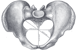
 Diameters of superior aperture of lesser pelvis -- female. (Pelvic brim is not labeled, but is identifiable as the central opening at the top.) | |
 Female pelvis. | |
| Identifiers | |
| FMA | 224780 |
| Anatomical terms of bone | |
The pelvic brim is the edge of the pelvic inlet. It is an approximately Mickey Mouse head-shaped line passing through the prominence of the sacrum, the arcuate and pectineal lines, and the upper margin of the pubic symphysis.
Structure
The pelvic brim is an approximately Mickey Mouse head-shaped line passing through the prominence of the sacrum, the arcuate and pectineal lines, and the upper margin of the pubic symphysis.
The pelvic brim is obtusely pointed in front, diverging on either side, and encroached upon behind by the projection forward of the promontory of the sacrum.
The oblique plane passing approximately through the pelvic brim divides the internal part of the pelvis (pelvic cavity) into the false or greater pelvis and the true or lesser pelvis. The false pelvis, which is above that plane, is sometimes considered to be a part of the abdominal cavity, rather than a part of the pelvic cavity. In this case, the pelvic cavity coincides with the true pelvis, which is below the above-mentioned plane.
The urinary bladder lies just above the anterior pelvic brim.[1] The sigmoid colon also passes close to the pelvic brim.[2]
Clinical significance
The pelvic brim may be a site of compression of structures that pass through the pelvic inlet. This can include the femoral nerves, which may occur during abdominal surgery.[3]
See also
Additional images
-
Pelvis
References
![]() This article incorporates text in the public domain from page 238 of the 20th edition of Gray's Anatomy (1918)
This article incorporates text in the public domain from page 238 of the 20th edition of Gray's Anatomy (1918)
- ^ Wilson, David A., ed. (2012-01-01), "Ultrasound: Urinary Tract", Clinical Veterinary Advisor, Saint Louis: W.B. Saunders, pp. 836–838, doi:10.1016/b978-1-4160-9979-6.00269-5, ISBN 978-1-4160-9979-6, retrieved 2021-01-30
- ^ Jacob, S. (2008-01-01), Jacob, S. (ed.), "Chapter 4 - Abdomen", Human Anatomy, Churchill Livingstone, pp. 71–123, doi:10.1016/b978-0-443-10373-5.50007-5, ISBN 978-0-443-10373-5, retrieved 2021-01-30
- ^ Lalkhen, Abdul Ghaaliq (2015-01-01), Tubbs, R. Shane; Rizk, Elias; Shoja, Mohammadali M.; Loukas, Marios (eds.), "Chapter 37 - Perioperative Peripheral Nerve Injuries Associated with Surgical Positioning", Nerves and Nerve Injuries, San Diego: Academic Press, pp. 587–602, ISBN 978-0-12-802653-3, retrieved 2021-01-30
External links
- Anatomy photo:44:os-0504 at the SUNY Downstate Medical Center

