
User:Rettootje/sandbox

Hypernasal Speech
Introduction[edit]
Rhinolalia = nasal speech, aperta = open, is the medical term for hypernasal speech. The other terms are hyperrhinolalia and open nasality. Hypernasality is a disorder of nasal speech when the sound of the voice is different, an abnormal resonance. There is an increased airflow through the nose during speech caused by open and nasal cavity due to incomplete closure of the soft palate and/or velopharyngeal sphincter. [1]
Anatomy[edit]

The palate exists of two components, the hard palate, palatum durum and the soft palate, palatum molle. The uvula is connected with the soft palate. This is the little thing in the back of the throat that comes up when one says ‘a’. The uvula can be lifted against the back throat wall to close the nasal cavity. This happens when you swallow or speak. In nasal sounds (‘m’, ‘n’ and ‘ng’) the uvula remains relaxed, thereby enabling the air to go through the nose.
The movements of the soft palate and the uvula are made possible by a sphincter: the velopharyngeal sphincter. Most children control this muscle around the age of three. Close to this velopharyngeal sphincter is the exit of the Eustachian tube. The Eustachian tube connects the middle ear and nasal pharynx. In a normal situation the tube ensures aeration and secretions drainage of the middle ear. The tube is very narrow and closed at rest. It opens in swallowing and yawning, controlled by the muscles of the soft palate. Children with a cleft palate have difficulties to control these muscles en with that their Eustachian tube. They are unable to open their Eustachian tube. The secretions of the ear will accumulate in the middle ear when the tube is dysfunctional for a longer time. This can cause hearing loss and middle ear infections. Ultimately, a hearing loss can lead to impaired speech and language development. [2] [3]
-
 Airflow mouth
 -
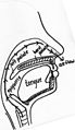 Airflow nose


Causes[edit]

The general term of the different disorders of the velopharyngeal valve is velopharyngeal dysfunction (VPD). This term includes three subterms:
- velopharyngeal insufficiency (VPI)
- velopharyngeal incompetence (VPI)
- velopharyngeal mislearning
Velopharyngeal insufficiency can be caused by an abnormality of the structures or anatomy of the throat. It occurs in children with a history of cleft palate or a submucous cleft. They have a short or abnormal velum. VPI can also be seen after adenoidectomy.
Velopharyngeal incompetence is a defective closure of the velopharyngeal valve due to a neurologic disorder or injury. This can be a cerebral palsy or traumatic brain injury. Some neurologic diseases have problems with the velopharyngeal sphincter. The sphincter does not work quickly and exactly enough.
Sometimes children seem to have no abnormalities but do have a hypernasal speech. A velopharyngeal mislearning indicates that the child has been imitating or has never learned how to use the valve correctly. For proper treatment a proper diagnosis is essential. [4][5] [6]
Diagnosis[edit]
There are several methods to diagnose hypernasality. First of all the speech therapist is listening to the child and analysis the perceptual speech while he/she is recording it. [7] [8] The child cannot say oral sounds, these are the vowels and the consonants. Only the nasal sounds can be said, these are the /m/, the /n/ and /ng/. [9] [10] Sometimes a hearing test is desirable.[11]
The mirror test is non-invasive and can easily evince the nasal air escape. The mirror is held beneath the nose while the child pronounces the vowels. The test is positive if the mirror is fogging.
A pressure-flow technique is an objective information for measuring velopharyngeal orifice area during the speech.
Another technique is a video nasopharyngeal endoscopy, which observes the velopharyngeal function, the movement of the soft palate and the back, both sides of the pharyngeal walls. It is a very small scope which will be placed in the back of the nasal cavity. The doctor will ask the child to say a few words. The patient has to be at least three or four years of age to undergo this last two techniques, as id needs a certain amount of co-operation.
The cinefluoroscopy gives dynamic visualisation and can easier be applied to younger children, but a great disadvantage is the radiation exposure. [12] [13]
The nasometer is an objective test. The patient wears a headset, where the oral and nasal cavities are separated by a plate. On both sides of the plate are microphones and give signals. With this technique the ration of the nasality can be calculated. It determines the size of the nasality. The higher the percentage the more nasality. [14]
Treatment[edit]
Speech therapist - In case of muscle weakness or cleft palate, special exercises can help to improve the muscle strength of the soft palate. These exercises are aimed at decreasing the airflow through the nose and thereby increasing intelligibility.
For good intelligibility it is necessary that the nasal cavity can be closed. All sounds, except for the ‘m’, ‘n’ and ‘ng’ sounds have an airflow only through the mouth. The muscles of the soft palate can raise it to close the nasal cavity. Normally a child can control these movements from the age of three. Children with a cleft palate often have difficulties with the muscles (not fully constructed or controllable) and are therefore unable to close the nasal cavity (completely).
Exercises[edit]
Blowing. If a child finds it difficult to blow, pinching the nose can help to regulate the airflow. Encourage the child to practise without pinching the nose.
- Blow out a candle
- Blowing away tissues with a drinking straw and with the mouth.
Sucking
- Drink milk with a drinking straw
This treatment is only useful if the deviations are small. Severe deviations should be treated surgically. [15]
Surgery[edit]
The two main surgical techniques for correcting the soft palate are the posterior pharyngeal flap and the sphincter pharyngoplasty.
Posterior pharyngeal flap This technique is mostly used for vertical clefts of the soft palate. The surgeon cuts through the upper layers of the back of the throat, creating a little square of tissue. This flap remains attached on one side, usually the upper side. The other side is attached to the (parts of) the soft palate. This ensures that the nasal cavity is partially separated from the oral cavity. When the child speaks, the remaining openings close from the side because of the narrowing of the throat caused by the muscle movements necessary for speaking. In relaxed state, the openings allow breathing through the nose.
Sphincter pharyngoplasty This technique is mostly used for horizontal clefts of the soft palate. Two small flaps are made on the left and right side of the entrance to the nasal cavity. They are attached to the back of the throat, thereby creating an opening exactly opposite to that of the posterior pharyngeal flap. To get a good result, patients must have a good palatal motion, as the occlusion of the nasal cavity is mainly done by the muscles already existing and functioning.
After surgical interventions, speech therapy is necessary to learn how to control the newly constructed flaps. [16]
| Rettootje/sandbox |
|---|
Complications[edit]
The most common complications of the posterior pharyngeal wall flap are hyponasality, nasal obstruction, snoring and sleep apnea. Rarer is a flap separation, sinusitis, postoperative bleeding and aspiration pneumonia. Complications of the sphincter pharyngoplasty are snoring, nasal obstruction, swallowing and difficulty with blowing nose. Both techniques have complications, however, some researches suggest that sphincter pharyngoplasty develops less hyponasality and obstructive sleep symptoms than the posterior pharyngeal wall flap. Both surgeries have a favourable effect on the function of the Eustachian tube. [17] [18] [19] [20]
References[edit]
- ^ http://projects.topshare.com/publicitem.m?key=kcts&pgid=28098&trail=/kcts/28093/28097/28098
- ^ http://www.schisis.nl/nvsca/pag.php?id=paK&item=pal#palsluit01
- ^ Stegenga,B. Vissink,A. Bont, L.G.M de. “Mondziekten & kaakchirurgie”. 2000, Van Gorcum & Comp. p 388
- ^ http://projects.topshare.com/publicitem.m?key=kcts&pgid=28098&trail=/kcts/28093/28097/28098
- ^ http://www.cincinnatichildrens.org/assets/0/78/759/781/65e90133-9243-4926-a065-8a97951944fb.pdf
- ^ http://emedicine.medscape.com/article/873018-overview#a0101
- ^ http://www.cpcjournal.org/doi/full/10.1597/1545-1569%282000%29037%3C0112%3APPFASP%3E2.3.CO%3B2
- ^ http://childrensnyp.org/mschony/otolaryngology-hypernasality.html
- ^ Gelder, van J. De open neusspraak. Ned.T.Geneesk. 1957. 101:1005-10
- ^ http://projects.topshare.com/publicitem.m?key=kcts&pgid=28098&trail=/kcts/28093/28097/28098
- ^ http://www.entcolumbia.org/hypernas.html
- ^ http://projects.topshare.com/publicitem.m?key=kcts&pgid=28098&trail=/kcts/28093/28097/28098
- ^ Probst,R. (2006) “Basic otorhinolaryngology: a step-by-step learning guide”. Stuttgart: Thieme. p.401
- ^ http://www.kayelemetrics.com/index.php?option=com_product&Itemid=3&controller=product&task=learn_more&cid%5B%5D=78
- ^ http://www.schisis.nl/nvsca/pag.php?id=paK&item=spraak
- ^ Lianne M. de Serres, Results with sphincter pharyngoplasty and pharyngeal flap. International Journal of Pediatric Otorhinolaryngology: April 1999, Vol. 48, Issue 1 , Pages 17-25
- ^ http://emedicine.medscape.com/article/873018-overview
- ^ Gerald M. Sloan (2000) Posterior Pharyngeal Flap and Sphincter Pharyngoplasty: The State of the Art. The Cleft Palate-Craniofacial Journal: February 2000, Vol. 37, No. 2, pp. 112-122.
- ^ Lianne M. de Serres, Results with sphincter pharyngoplasty and pharyngeal flap. International Journal of Pediatric Otorhinolaryngology: April 1999, Vol. 48, Issue 1 , Pages 17-25
- ^ P.H.M. Spauwen, De invloed van chirurgische behandeling van open neusspraak op horen en spreken. Ned Tijdschr Geneeskd. 1987;131:161-6