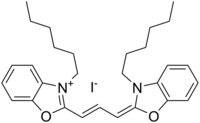
DiOC6
Appearance

| |
| Names | |
|---|---|
| IUPAC name
3-Hexyl-2-[3-(3-hexyl-2(3H)benzoxazolylidene)-1-propenyl]benzoxazolium iodide
| |
| Other names
3,3′-Dihexyloxacarbocyanine iodide
| |
| Identifiers | |
3D model (JSmol)
|
|
| ChemSpider | |
| ECHA InfoCard | 100.155.178 |
PubChem CID
|
|
| UNII | |
CompTox Dashboard (EPA)
|
|
| |
| |
| Properties | |
| C29H37IN2O2 | |
| Molar mass | 572.531 g·mol−1 |
Except where otherwise noted, data are given for materials in their standard state (at 25 °C [77 °F], 100 kPa).
| |
DiOC6 (3,3′-dihexyloxacarbocyanine iodide) is a fluorescent dye used for the staining of a cell's endoplasmic reticulum,[2] vesicle membranes and mitochondria.[3] Binding to these structures occurs via the dye's hydrophilic groups. DiOC6 can be used to label living cells, however they are quickly damaged due to the dye's extreme phototoxicity, so cells stained with this dye can only be exposed to light for short periods of time. When exposed to blue light, the dye fluoresces green.
See also
[edit]References
[edit]- ^ 3,3′-Dihexyloxacarbocyanine iodide at Sigma-Aldrich
- ^ Terasaki M. (1989). "Fluorescent labeling of endoplasmic reticulum". Methods Cell Biol. 29: 125–35. doi:10.1016/s0091-679x(08)60191-0. PMID 2643757.
- ^ Koning AJ; et al. (1993). "DiOC6 staining reveals organelle structure and dynamics in living yeast cells". Cell Motil Cytoskeleton. 25 (2): 111–28. doi:10.1002/cm.970250202. PMID 7686821.
