Adductor hiatus
| Adductor hiatus | |
|---|---|
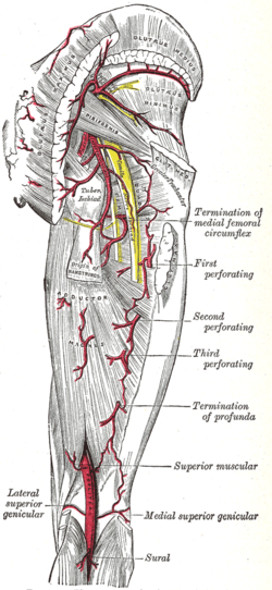
 The arteries of the gluteal and posterior femoral regions. (Adductor hiatus is not labeled, but popliteal artery is visible at bottom center.) | |
 Deep muscles of the medial femoral region. (Adductor hiatus visible as hole in adductor magnus at lower left.) | |
| Details | |
| Identifiers | |
| Latin | hiatus adductorius |
| TA98 | A04.7.03.008 |
| TA2 | 2634 |
| FMA | 58784 |
| Anatomical terminology | |
The adductor hiatus is a gap between the adductor magnus muscle and the femur that allows the passage of the femoral vessels from the anterior thigh to the posterior thigh and then the popliteal fossa. It is the termination of the adductor canal and lies about 2 inches superior to the knee.
Four structures are associated with the adductor hiatus. However, only two structures enter and then leave through the hiatus; namely the femoral artery and femoral vein. Those vessels become the popliteal vessels (popliteal artery and popliteal vein) immediately after they leave the hiatus, where they form a network of anastomoses called the genicular vessels. The genicular vessels supply the knee joint.
The other two structures that are associated with the adductor hiatus are the saphenous branch of descending genicular artery and the saphenous nerve. The saphenous nerve doesn't actually leave through the adductor hiatus but penetrates superficially half way through the adductor canal.
Clinical considerations
A fracture of the femur just superior to the knee joint will most likely affect the adductor hiatus and may cause impairment of the blood supply to the lower leg.
Additional images
-
Schema of the arteries arising from the external iliac and femoral arteries.

