
File:Cranial endocast of Baryonyx.jpg

Original file (2,764 × 3,735 pixels, file size: 2.25 MB, MIME type: image/jpeg)
| This is a file from the Wikimedia Commons. Information from its description page there is shown below. Commons is a freely licensed media file repository. You can help. |
Summary
| Description |
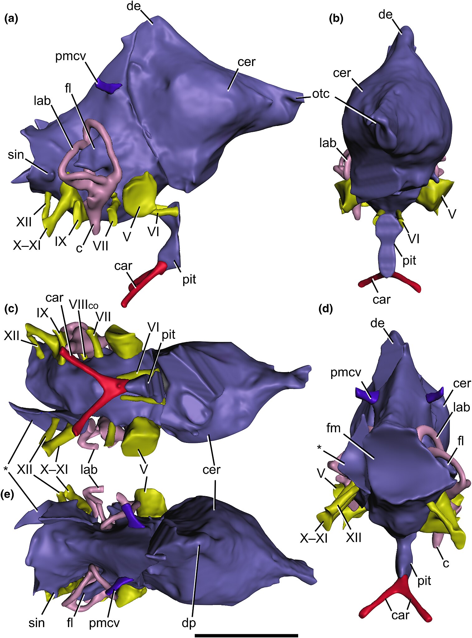
Cranial endocast of Baryonyx walkeri (NHMUK PV R9951), reconstructed from CT scans, in (a) right lateral, (b) anterior, (c) ventral, (d) posterior and (e) dorsal views. Vascular structures and endosseous labyrinth also depicted. Abbreviations: c, cochlea; car, cerebral internal carotid artery canal; cer, cerebral hemisphere; de, dorsal expansion; fl, floccular lobe; lab, endosseous labyrinth; otc, olfactory tract; pit, pituitary; pmcv, posterior middle cerebral vein canal; sin, blind dural venous sinus of the hindbrain; V, trigeminal nerve canal; VI, abducens nerve canal; VII, facial nerve canal, VIIIco, cochlear ramus of the vestibulocochlear nerve; IX, glossopharyngeal nerve canal; X–XI, shared canal for the vagus, and accessory nerves, and accompanying vessels; XII, hypoglossal nerve canal. Asterisk (*) marks the disarticulated left otoccipital portion of the endocast. Scale bar: 50 mm. |
| Date | |
| Source | https://onlinelibrary.wiley.com/doi/10.1111/joa.13837 |
| Author | Chris Tijani Barker, Darren Naish, Jacob Trend, Lysanne Veerle Michels, Lawrence Witmer, Ryan Ridgley, Katy Rankin, Claire E. Clarkin, Philipp Schneider, Neil J. Gostling |
Licensing
- You are free:
- to share – to copy, distribute and transmit the work
- to remix – to adapt the work
- Under the following conditions:
- attribution – You must give appropriate credit, provide a link to the license, and indicate if changes were made. You may do so in any reasonable manner, but not in any way that suggests the licensor endorses you or your use.
File history
Click on a date/time to view the file as it appeared at that time.
| Date/Time | Thumbnail | Dimensions | User | Comment | |
|---|---|---|---|---|---|
| current | 20:15, 5 May 2023 | | 2,764 × 3,735 (2.25 MB) | FunkMonk | {{Information |description=Cranial endocast of Baryonyx walkeri (NHMUK PV R9951), reconstructed from CT scans, in (a) right lateral, (b) anterior, (c) ventral, (d) posterior and (e) dorsal views. Vascular structures and endosseous labyrinth also depicted. Abbreviations: c, cochlea; car, cerebral internal carotid artery canal; cer, cerebral hemisphere; de, dorsal expansion; fl, floccular lobe; lab, endosseous labyrinth; otc, olfactory tract; pit, pituitary; pmcv, posterior middle cerebral vein... |
File usage
The following page uses this file: