Ilium (bone)
| Ilium of pelvis | |
|---|---|
 Overview of Ilium as largest region of the pelvis. | |
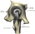
 Capsule of hip-joint (distended). Posterior aspect. (Ilium labeled at top.) | |
| Details | |
| Identifiers | |
| Latin | Os ilium |
| MeSH | D007085 |
| TA98 | A02.5.01.101 |
| TA2 | 1317 |
| FMA | 16589 |
| Anatomical terms of bone | |
The ilium (/ˈɪlɪəm/) is the uppermost and largest region of the coxal bone, and appears in most vertebrates including mammals and birds, but not bony fish. All reptiles have an ilium except snakes, although some snake species have a tiny bone which is considered to be an ilium.[1]
The name comes from the Latin (ile, ilis), meaning "groin" or "flank."[2]
The ilium of the human is divisible into two parts, the body and the ala; the separation is indicated on the top surface by a curved line, the arcuate line, and on the external surface by the margin of the acetabulum.
Structure
Body (corpus ossis ilium)
The body enters into the formation of the acetabulum, of which it forms rather less than two-fifths. Its external surface is partly articular, partly non-articular; the articular segment forms part of the lunate surface of the acetabulum, the non-articular portion contributes to the acetabular fossa.
The internal surface of the body is part of the wall of the lesser pelvis and gives origin to some fibers of the obturator internus.
Below, it is continuous with the pelvic surfaces of the ischium and pubis, with only a faint line indicating the place of union.
The body of ilium together with the wing forms the ilium.
Ala (ala ossis ilium)
The wing of ilium (or ala) is the large expanded portion which bounds the greater pelvis laterally. It presents for examination two surfaces—an external and an internal—a crest, and two borders—an anterior and a posterior.
Development
This section needs expansion. You can help by adding to it. (November 2013) |
In other vertebrates
Dinosaurs
The clade Dinosauria is divided into the Saurischia and Ornithischia based on hip structure, including importantly that of the ilium.[3] In both Saurischians and Ornithischians, the ilium extends laterally to both sides from the axis of the body. The other two hip bones, the ischium and the pubis, extend ventrally down from the ilium towards the belly of the animal. The acetabulum, which can be thought of as a "hip-socket", is an opening on each side of the pelvic girdle formed where the ischium, ilium, and pubis all meet, and into which the head of the femur inserts. The orientation and position of the acetabulum is one of the main morphological traits that caused dinosaurs to walk in an upright posture with their legs directly underneath their bodies. The brevis fossa is a deep groove in the underside of the postacetabular process, the rear part of the ilium. The brevis shelf is the bony ridge at the inner side of the fossa, the bone wall forming the internal face of the rear part of the ilium, which functions as an attachment area for a tail muscle, the musculus caudofemoralis brevis.[4] Often, close to the hip-socket the lower edge of the outer face of the postacetabular process is positioned higher than the edge of the brevis shelf, exposing the latter in side view. Some dinosaurs did not have lliums such as the widely known pteradactal.
-
Ornithischian pelvic structure (left side)
-
Saurischian pelvic structure (left side).
Clinical significance
This section needs expansion. You can help by adding to it. (November 2013) |
Biiliac width
In humans, biiliac width is an anatomical term referring to the widest measure of the pelvis between the outer edges of the upper iliac bones.
Biiliac width has the following common synonyms: pelvic bone width, biiliac breadth, intercristal breadth/width, bi-iliac breadth/width and biiliocristal breadth/width.
It is best measured by anthropometric calipers (an anthropometer designed for such measurement is called a pelvimeter). Attempting to measure biiliac width with a tape measure along a curved surface is inaccurate.
The biiliac width measure is helpful in obstetrics because a pelvis that is significantly too small or too large can have obstetrical complications. For example, a large baby or a small pelvis often lead to death unless a caesarean section is performed.[5]
It is also used by anthropologists to estimate body mass.[6]
History
The 'English' name ilium as bone of the pelvis can be traced back to the writings of anatomists Andreas Vesalius, whom coined the expression os ilium.[7] In this expression ilium can be considered as the genitive plural of the nominative singular of the noun ile.[7] Ile in classical Latin can refer to the flank of the body,[8] or to the groin,[8] or the part of the abdomen from the lowest ribs to the pubes.[8] Ile is usually encountered as plural (ilia) in classical Latin.[8] The os ilium can literally be translated as bone (Latin: os[8] ) of the flanks.
More than a millennium earlier the ossa ilium were described by the Greek physician Galen, and referred to as, with a quite similar expression, τά πλατέα λαγόνων ὀστᾶ, the flat bones of the flanks,[7] with λαγών for flank.[9] In anatomic Latin, the expression os lagonicum[10] can also be found, based on Ancient Greek λαγών. In modern Greek the nominalized adjective λαγόνιο[11] is used to refer to the os ilium.
In Latin and Greek it is not uncommon to nominalize adjectives, e.g. stimulantia from remedia stimulantia[12] or ὁ ἐγκέφαλος from ὁ ἐγκέφαλος μυελός.[13] The name ilium as used in English[14][15] can not be considered as nominalized adjective derived from the full Latin expression os ilium, as ilium in this expression is a genitive plural of a noun[7] and not a nominative singular of an adjective. The form ilium in English is however thought to be derived from the Latin word ilium,[16] an orthographic variant in Latin of ile,[8][16] flank or groin.[8] Whereas the expression of Andreas Vesalius os ilium appropriately expresses bone of the flanks, the sole term ilium as used in English, lacks this precision and has to be literally translated as groin or flank.
There exists however in classical Latin an adjective ilius/ilia/ilium. This adjective however means not with respect to the flanks, but Trojan.[8] Troy is referred to in classical Latin as Ilium,[8] Ilion[8] or Ilios[17] and in ancient Greek as Ἴλιον[9] or Ἴλιος.[9]
The first editions of the official Latin nomenclature, Nomina Anatomica of the first 80 years (first in 1895) used the Vesalian expression os ilium.[18][19][20][21][22][23] In the subsequent editions from 1983[24] and 1989[25] the expression os ilium was altered to os ilii. This latter expression supposes a genitive singular of the alternate noun ilium instead of a genitive plural of the noun ile. Quite inconsistently, in the 1983 edition[24] of the Nomina Anatomica the genitive plural of ile (instead of ilium) is still being used in such expressions as vena circumflexa ilium superficialis. In the current 1998 edition of the Nomina Anatomica, rebaptized as Terminologia Anatomica the expression os ilium is reintroduced and os ilii deleted.
Additional images
-
Pelvic girdle
-
Male pelvis.
-
Right hip bone. Internal surface.
-
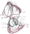
Right hip bone. External surface. (Body of ilium is the top of the blue circle in the center, and the wing of the ilium is the portion above that. Crest of ilium is labeled at top.)
-
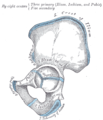
Plan of ossification of the hip bone.
-
Left hip-joint, opened by removing the floor of the acetabulum from within the pelvis.
-
Pelvis (French labels)
-
Pelvis
See also
References
![]() This article incorporates text in the public domain from page 236 of the 20th edition of Gray's Anatomy (1918)
This article incorporates text in the public domain from page 236 of the 20th edition of Gray's Anatomy (1918)
- ^ Jacobson, Elliott R. (2007). Infectious Diseases and Pathology of Reptiles. CRC Press. p. 7. ISBN 0-8493-2321-5. ISBN 9780849323218. Retrieved 2009-01-09.
- ^ Taber, Clarence Wilbur; Venes, Donald (2005). Taber's cyclopedic medical dictionary. Philadelphia: F.A. Davis. ISBN 0-8036-1207-9.
- ^ Seeley, H.G. (1888). "On the classification of the fossil animals commonly named Dinosauria." Proceedings of the Royal Society of London, 43: 165-171.
- ^ Martin, A.J. (2006). Introduction to the Study of Dinosaurs. Second Edition. Oxford, Blackwell Publishing. pg. 299-300. ISBN 1-4051-3413-5.
- ^ "Encyclopedia of Medicine: Cesarean Section". eNotes.
- ^ Ruff C, Niskanenb M, Junnob J, Jamisonc P (2005). "Body mass prediction from stature and bi-iliac breadth in two high latitude populations, with application to earlier higher latitude humans" (PDF). Journal of Human Evolution. 48 (4): 381–392. doi:10.1016/j.jhevol.2004.11.009. PMID 15788184. Retrieved 2006-07-26.
- ^ a b c d Hyrtl, J. (1880). Onomatologia Anatomica. Geschichte und Kritik der anatomischen Sprache der Gegenwart. Wien: Wilhelm Braumüller. K.K. Hof- und Universitätsbuchhändler.
- ^ a b c d e f g h i j Lewis, C.T. & Short, C. (1879). A Latin dictionary founded on Andrews' edition of Freund's Latin dictionary. Oxford: Clarendon Press.
- ^ a b c Liddell, H.G. & Scott, R. (1940). A Greek-English Lexicon. revised and augmented throughout by Sir Henry Stuart Jones. with the assistance of. Roderick McKenzie. Oxford: Clarendon Press.
- ^ Kossmann, R. (1895). Die gynäcologische Anatomie und ihre zu Basel festgestellte Nomenclatur. Monatsschrift für Geburtshülfe und Gynaekologie, 2 (6), 447-472.
- ^ Schleifer, S.K. (Ed.) (2011). Corpus humanum, The human body, Le corps humain, Der menschliche Körper, Il corpo umano, El cuerpo humano, Ciało człowieka, Människokroppen, Menneskekroppen, Τό ανθρώπινο σῶμα, ЧЕЛОВЕК. FKG.
- ^ Arnaudov, G.D. (1964). Terminologia medica polyglotta. Latinum-Bulgarski-Russkij-English-Français-Deutsch. Sofia: Editio medicina et physcultura.
- ^ Kraus, L.A. (1844). Kritisch-etymologisches medicinisches Lexikon (Dritte Auflage). Göttingen: Verlag der Deuerlich- und Dieterichschen Buchhandlung.
- ^ Dorland, W.A.N. & Miller, E.C.L. (1948). The American illustrated medical dictionary.’’ (21st edition). Philadelphia/London: W.B. Saunders Company.
- ^ Dirckx, J.H. (Ed.) (1997).Stedman’s concise medical dictionary for the health professions. (3rd edition). Baltimore: Williams & Wilkins.
- ^ a b Klein, E. (1971). A comprehensive etymological dictionary of the English language. Dealing with the origin of words and their sense development thus illustration the history of civilization and culture. Amsterdam: Elsevier Science B.V.
- ^ Wageningen, J. van & Muller, F. (1921). Latijnsch woordenboek. (3de druk). Groningen/Den Haag: J.B. Wolters’ Uitgevers-Maatschappij
- ^ His, W. (1895). Die anatomische Nomenclatur. Nomina Anatomica. Der von der Anatomischen Gesellschaft auf ihrer IX. Versammlung in Basel angenommenen Namen. Leipzig: Verlag Veit & Comp.
- ^ Kopsch, F. (1941). Die Nomina anatomica des Jahres 1895 (B.N.A.) nach der Buchstabenreihe geordnet und gegenübergestellt den Nomina anatomica des Jahres 1935 (I.N.A.) (3. Auflage). Leipzig: Georg Thieme Verlag.
- ^ Stieve, H. (1949). Nomina Anatomica. Zusammengestellt von der im Jahre 1923 gewählten Nomenklatur-Kommission, unter Berücksichtigung der Vorschläge der Mitglieder der Anatomischen Gesellschaft, der Anatomical Society of Great Britain and Ireland, sowie der American Association of Anatomists, überprüft und durch Beschluß der Anatomischen Gesellschaft auf der Tagung in Jena 1935 endgúltig angenommen. (4th edition). Jena: Verlag Gustav Fischer.
- ^ Donáth, T. & Crawford, G.C.N. (1969). Anatomical dictionary with nomenclature and explanatory notes. Oxford/London/Edinburgh/New York/Toronto/Syney/Paris/Braunschweig: Pergamon Press.
- ^ International Anatomical Nomenclature Committee (1966). Nomina Anatomica. Amsterdam: Excerpta Medica Foundation.
- ^ International Anatomical Nomenclature Committee (1977). Nomina Anatomica, together with Nomina Histologica and Nomina Embryologica. Amsterdam-Oxford: Excerpta Medica.
- ^ a b International Anatomical Nomenclature Committee (1983). Nomina Anatomica, together with Nomina Histologica and Nomina Embryologica. Baltimore/London: Williams & Wilkins
- ^ International Anatomical Nomenclature Committee (1989). Nomina Anatomica, together with Nomina Histologica and Nomina Embryologica. Edinburgh: Churchill Livingstone.
External links
- Anatomy photo:44:st-0701 at the SUNY Downstate Medical Center
- pelvis at The Anatomy Lesson by Wesley Norman (Georgetown University)


