
Micrograph
This article needs additional citations for verification. (September 2014) |


A micrograph or photomicrograph is a photograph or digital image taken through a microscope or similar device to show a magnified image of an item. This is opposed to a macrographic image, which is at a scale that is visible to the naked eye.
Neuropathologist Solomon C. Fuller designed and created the first photomicrograph in[1] 1900.
Micrographs are widely used in all fields of microscopy.
Types
Photomicrograph
A light micrograph or photomicrograph is a micrograph prepared using an optical microscope, a process referred to as photomicroscopy. At a basic level, photomicroscopy may be performed simply by hooking up a regular camera to a microscope, thereby enabling the user to take photographs at reasonably high magnification.
Roman Vishniac was a pioneer in the field of photomicroscopy, specializing in the photography of living creatures in full motion. He also made major developments in light-interruption photography and color photomicroscopy.
Electron micrograph
An electron micrograph is a micrograph prepared using an electron microscope. However, the term electron micrograph is not used in electron microscopy. Common designation is a micrograph.
Digital micrography
Digital micrography is a digital picture obtained either directly with a microscope or by scanning of a photomicrograph. The terms usage is somewhat confusing, since today photo usually means digital photography anyway.[citation needed] Digital micrographs are now commonly obtained using a USB microscope attached directly to a home computer or laptop.
Today, an add-on three-in-one macro lens which has capability to take wide-angle, fish-eye and macro with 7x, 14x and 21x magnification can be attached on iPhone5/5s or 5th generation of iPod.[2]
Magnification and micron bars
Micrographs usually have micron bars, or magnification ratios, or both.
Magnification is a ratio between size of object on a picture and its real size. Unfortunately, magnification is somewhat a misleading parameter. It depends on a final size of a printed picture, and therefore varies with variation in picture size. Editors of journals and magazines routinely resize a figure to fit the page, making any magnification number provided in the figure legend incorrect. A scale bar, or micron bar, is a bar of known length displayed on a picture. The bar can be used for measurements on a picture. When a picture is resized a bar is also resized. If a picture has a bar, the magnification can be easily calculated. Ideally, all pictures destined for publication/presentation should be supplied with a scale bar; the magnification ratio is optional. All but one (limestone) of the micrographs presented on this page do not have a micron bar; supplied magnification ratios are likely incorrect, as they were not calculated for pictures at the present size.
Gallery
-
 Measurements of a large Colpodium at 400x.
 -
 Measurements of a large amoeba at 400x.
 -
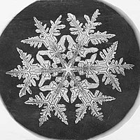 Snowflake micrograph by Wilson Bentley, 1890


See also
References
- ^ "Fuller, Jr., Solomon Carter 1872–1953 Research". Encyclopedia.com. N. p., 2016. Web. 13 Mar. 2016.
- ^ "Olloclip debuts Macro 3-in-1 lens for iPhone and iPod touch (hands-on)". Retrieved December 5, 2013.
External links
- Make a Micrograph – This presentation by the research department of Children's Hospital Boston shows how researchers create a three-color micrograph.
- Shots with a Microscope – a basic, comprehensive guide to photomicrography
- Scientific photomicrographs – free scientific quality photomicrographs by Doc. RNDr. Josef Reischig, CSc.
- Micrographs of 18 natural fibres by the International Year of Natural Fibres 2009
- Seeing Beyond the Human Eye Video produced by Off Book (web series)
- [1] - Solomon C. Fuller bio