
Inferior vena cava
| Inferior vena cava | |
|---|---|
 Superior vena cava, inferior vena cava, azygos vein and their tributaries. | |
| Details | |
| Source | common iliac vein lumbar veins testicular vein renal vein suprarenal vein hepatic vein |
| Drains to | heart |
| Artery | abdominal aorta |
| Identifiers | |
| Latin | vena cava inferior |
| MeSH | D014682 |
| TA98 | A12.3.09.001 |
| TA2 | 4991 |
| FMA | 10951 |
| Anatomical terminology | |
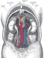
The inferior vena cava (or IVC), also known as the posterior vena cava,[1] is the large vein that carries de-oxygenated blood from the lower half of the body into the right atrium of the heart.
It is posterior to the abdominal cavity and runs alongside of the vertebral column on its right side (i.e. it is a retroperitoneal structure). It enters the right atrium at the lower right, back side of the heart.
Drainage patterns
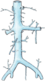
The IVC is formed by the joining of the left and right common iliac veins and brings blood into the right atrium of the heart. It also anastomoses with the azygos vein system (which runs on the right side of the vertebral column) and venous plexuses next to the spinal cord.
The caval opening is at T8. The specific levels of the tributaries are as follows:
| Vein | Level |
| hepatic veins | T8 |
| inferior phrenic vein | T8 |
| suprarenal vein | L1 |
| renal veins | L1 |
| gonadal vein | L2 |
| lumbar veins | L1-L5 |
| common iliac veins | L5 |
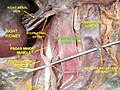
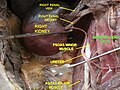
Because the IVC is not centrally located, there are some asymmetries in drainage patterns. The gonadal veins and suprarenal veins drain into the IVC on the right side, but into the renal vein on the left side, which in turn drains into the IVC. By contrast, all the lumbar veins and hepatic veins usually drain directly into the IVC.
The tributaries of Inferior vena cava can be remembered using the mnemonic, "I Like To Rise So High", for Illiac vein (common), Lumbar vein, Testicular vein, Renal vein, Suprarenal vein and Hepatic vein.[2]
Note that the vein that carries de-oxygenated blood from the upper half of the body is the superior vena cava.
Pathologies associated with the IVC
Health problems attributed to the IVC are most often associated with it being compressed (ruptures are rare because it has a low intraluminal pressure). Typical sources of external pressure are an enlarged aorta (abdominal aortic aneurysm), the gravid uterus (aortocaval compression syndrome) and abdominal maligancies, such as colorectal cancer, renal cell carcinoma and ovarian cancer. Since the inferior vena cava is primarily a right-sided structure, unconscious pregnant females should be turned on to their left side (the recovery position), to relieve pressure on it and facilitate venous return. In rare cases, straining associated with defecation can lead to restricted blood flow through the IVC and result in syncope (fainting).[3]
Occlusion of the IVC is rare, but considered life-threatening and is an emergency. It is associated with deep vein thrombosis, IVC filters, liver transplantation and instrumentation (e.g. catheter in the femoral vein).[4]
Embryology
In the embryo, the IVC and right atrium are separated by the Eustachian valve, also known in Latin as the valvula venae cavae inferioris (valve of the inferior vena cava). In the adult, this structure typically has totally regressed or remains as a small endocardial fold.[5]
Additional images
-
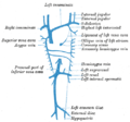 Diagram showing completion of development of the parietal veins.
 -
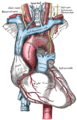
 Base and diaphragmatic surface of heart.
 -
 The arch of the aorta, and its branches.
 -
 The abdominal aorta and its branches.
 -
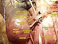
 Inferior vena cava
 -
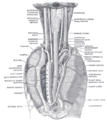 The position and relation of the esophagus in the cervical region and in the posterior mediastinum. Seen from behind.
 -
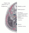 Transverse section of human embryo eight and a half to nine weeks old.
 -
 Human kidneys viewed from behind with spine removed
 -
 Liver and gallbladder
 -
 Inferior vena cava
 -
 Inferior vena cava
 -
 Inferior vena cava
 -
 Inferior vena cava
 -
 Inferior vena cava


See also
References
- ^ Organisms and Populations 3rd Ed.
- ^ "Medical mnemonic for IVC tributaries". LifeHugger. Retrieved 2009-12-16.
- ^ Brophy, CM; Evans, L; Sumpio, BE (1993). "Defecation syncope secondary to functional inferior vena caval obstruction during a Valsalva maneuver". Annals of vascular surgery. 7 (4): 374–7. doi:10.1007/BF02002893. PMID 8268080.
- ^ Geehan DM, Inferior Vena Caval Thrombosis, emedicine.com, URL: http://www.emedicine.com/med/topic2718.htm, Accessed: August 3, 2005.
- ^ Turhan Yavuz; Nazli, C; Kinay, O; Kutsal, A (2002). "Giant Eustachian Valve: with Echocardiographic Appearance of Divided Right Atrium". Texas Heart Institute Journal. 29 (4): 336–8. PMC 140300. PMID 12484622.
External links
- synd/1462 at Who Named It? - "Eustachian valve"
- Anatomy photo:40:13-0101 at the SUNY Downstate Medical Center - "Posterior Abdominal Wall: Tributaries to the Inferior Vena Cava"
- Anatomy image:8827 at the SUNY Downstate Medical Center
- Cross section image: pembody/body12a—Plastination Laboratory at the Medical University of Vienna