Inguinal lymph nodes
It has been suggested that Deep inguinal lymph nodes be merged into this article. (Discuss) Proposed since April 2017. |
| Inguinal lymph nodes | |
|---|---|

| |
 The lymph glands and lymphatic vessels of the lower extremity. | |
| Details | |
| Drains from | most of perineal region |
| Identifiers | |
| Latin | nodi lymphoidei inguinales superficiales |
| TA98 | A13.3.05.002 |
| FMA | 44226 |
| Anatomical terminology | |
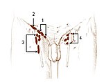
Inguinal lymph nodes are the lymph nodes in the inguinal region. They are found in the femoral triangle, and are grouped into superficial nodes, and deep nodes.
Superficial inguinal lymph nodes
- The superficial inguinal lymph nodes are divided into three groups:
- inferior – inferior of the saphenous opening of the leg, receive drainage from lower legs
- superolateral – on the side of the saphenous opening, receive drainage from the side buttocks and the lower abdominal wall.
- superomedial – located at the middle of the saphenous opening, take drainage from the perineum and outer genitalia.[1]
Deep inguinal lymph nodes
The deep inguinal lymph nodes:
Lymph node size
The mean size of an inguinal lymph node, as measured over the short-axis, is approximately 5.4 mm (range 2.1-13.6 mm), with two standard deviations above the mean being 8.8 mm.[3] A size of up to 10 mm is generally regarded as a cut-off value for normal vs abnormal inguinal lymph node size.[4]
Additional images
-
A view of the different inguinal lymph nodes
-
Murine inguinal lymph node beneath the bifurcation of superior epigastric vein. Bright structure visualised by MHC II-GFP construct, is the lymph node
References
- ^ "Superficial Inguinal Lymph Nodes -- Medical Definition". www.medilexicon.com. Retrieved 2016-05-09.
- ^ "lymph nodes and nerves". www.oganatomy.org. Retrieved 2016-05-09.
- ^ Bontumasi, Nicholas; Jacobson, Jon A.; Caoili, Elaine; Brandon, Catherine; Kim, Sung Moon; Jamadar, David (2014). "Inguinal lymph nodes: size, number, and other characteristics in asymptomatic patients by CT". Surgical and Radiologic Anatomy. 36 (10): 1051–1055. doi:10.1007/s00276-014-1255-0. ISSN 0930-1038.
- ^ Maha Torabi, MD;, Suzanne L. Aquino; and Mukesh G. Harisinghani (2004-09-01). "Current Concepts in Lymph Node Imaging". J Nucl Med. 45 (9): 1509–1518.
{{cite journal}}: CS1 maint: multiple names: authors list (link)


