
Otic vesicle
| Otic vesicle | |
|---|---|
 Embryo between eighteen and twenty-one days. | |
 Section through hind-brain and auditory vesicles of an embryo more advanced than that of Fig. 898. | |
| Details | |
| Precursor | auditory pit |
| Identifiers | |
| Latin | vesicula otica |
| TE | vesicle_by_E5.15.1.0.0.0.4 E5.15.1.0.0.0.4 |
| Anatomical terminology | |
When the mouth of the auditory pit is closed, and thus a shut sac, the auditory vesicle (otocyst or otic vesicle[1]), is formed; from it the epithelial lining of the membranous labyrinth is derived.
The vesicle becomes pear-shaped, and the neck of the flask is obliterated.
From the vesicle certain diverticula are given off which form the various parts of the membranous labyrinth.
One from the middle part forms the ductus and saccus endolymphaticus, another from the anterior end gradually elongates, and, forming a tube coiled on itself, becomes the cochlear duct, the vestibular extremity of which is subsequently constricted to form the canalis reuniens.
Three others appear as disk-like evaginations on the surface of the vesicle; the central parts of the walls of the disks coalesce and disappear, while the peripheral portions persist to form the semicircular ducts; of these the superior is the first and the lateral the last to be completed.
The central part of the vesicle represents the membranous vestibule, and is subdivided by a constriction into a smaller ventral part, the saccule, and a larger dorsal and posterior part, the utricle.
This subdivision is effected by a fold which extends deeply into the proximal part of the ductus endolymphaticus, with the result that the utricle and saccule ultimately communicate with each other by means of a Y-shaped canal.
The saccule opens into the cochlear duct, through the canalis reuniens, and the semicircular ducts communicate with the utricle.
Additional images
-
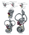 Lateral views of membranous labyrinth and acoustic complex. X 25 dia.
 -
 Median views of membranous labyrinth and acoustic complex in human embryos. X 25 dia.


References
- ^ ear-005b—Embryo Images at University of North Carolina
This neuroscience article is a stub. You can help Wikipedia by expanding it. |
This developmental biology article is a stub. You can help Wikipedia by expanding it. |