Anterior cruciate ligament: Difference between revisions
m Reverted edits by 140.211.123.153 (talk) to last version by 24.215.2.203 |
|||
| Line 32: | Line 32: | ||
The [[Lachman test]] is supported by most authorities to be the |
The [[Lachman test]] is supported by most authorities to be the |
||
most reliable and sensitive maneuver for the diagnosis of an ACL tear. |
most reliable and sensitive maneuver for the diagnosis of an ACL tear. |
||
By Resaish |
|||
==References== |
==References== |
||
Revision as of 17:07, 18 October 2007
| Anterior cruciate ligament | |
|---|---|
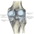
 Diagram of the right knee. (Anterior cruciate ligament labeled at center left.) | |
| Details | |
| From | lateral condyle of the femur |
| To | intercondyloid eminence of the tibia |
| Identifiers | |
| Latin | ligamentum cruciatum anterius |
| MeSH | D016118 |
| TA98 | A03.6.08.007 |
| TA2 | 1890 |
| FMA | 44614 |
| Anatomical terminology | |
The anterior cruciate ligament (or ACL) is one of the four major ligaments of the knee.
It connects from a posterio-lateral part of the femur to an anterio-medial part of the tibia. These attachments allow it to resist forces pushing the tibia forward relative to the femur.
More specifically, it is attached to the depression in front of the intercondyloid eminence of the tibia, being blended with the anterior extremity of the lateral meniscus.
It passes up, backward, and laterally, and is fixed into the medial and back part of the lateral condyle of the femur.
Injury
Although there are many ACL injuries, the ACL is next to the most commonly injured knee ligament, the posterior cruciate ligament (PCL)[1] and commonly injured by athletes. The ACL is often torn during sudden dislocation, tortion, or hyperextension of the knee. It is a very common injury in hockey, skiing, skating, basketball, and football due to the enormous amount of pressure, weight and number of blows the knee must withstand.
Diagnosis
The Lachman test is supported by most authorities to be the most reliable and sensitive maneuver for the diagnosis of an ACL tear.
By Resaish
References
See also
- Knee
- Lateral collateral ligament
- Medial collateral ligament
- Posterior cruciate ligament
- Anterior cruciate ligament reconstruction
Additional images
-
Right knee-joint, from the front, showing interior ligaments.
-
Left knee-joint from behind, showing interior ligaments.
-
Head of right tibia seen from above, showing menisci and attachments of ligaments.
-
Capsule of right knee-joint (distended). Posterior aspect.
External links
- Anatomy photo:17:02-0701 at the SUNY Downstate Medical Center - "Major Joints of the Lower Extremity: Knee Joint"
- Anatomy figure: 17:07-08 at Human Anatomy Online, SUNY Downstate Medical Center - "Superior view of the tibia."
- Anatomy figure: 17:08-03 at Human Anatomy Online, SUNY Downstate Medical Center - "Medial and lateral views of the knee joint and cruciate ligaments."
- Template:EMedicineDictionary
- MedicalMnemonics.com: 2081


