Renal biopsy: Difference between revisions
Updated intro |
→Complications: Updated. More comprehensive and realistic. |
||
| Line 53: | Line 53: | ||
==Complications== |
==Complications== |
||
Serious complications of renal biopsy are |
Serious complications of renal biopsy are uncommon. |
||
Bleeding is the most common complication of renal biopsy. Rarely, bleeding is severe enough to require a [[blood transfusion]] or surgery. Most patients who undergo renal biopsy notice [[Hematuria|blood in the urine]] for several days after the procedure. Patients with urine that is bright red or brown for longer than one week should consult with their healthcare provider. |
|||
Pain is a common problem, although it is usually mild to moderate and resolves within a few hours. Medications can be given to reduce pain after the procedure. Patients who experience severe or prolonged pain should notify their healthcare provider; this can be a sign of a [[Thrombus|blood clot]] that is obstructing the ureter (tube that leads to the bladder) or a large hematoma (a mass of clotted blood) that stretches the kidney. |
|||
The most common complication of kidney biopsy is bleeding. This reflects the density of blood vessels within the kidney and observation that individuals with [[kidney failure]] take longer to stop bleeding after trauma ([[uraemic coagulopathy]]). Bleeding complications include a collection of blood adjacent to or around the kidney ([[perinephric haematoma]]), bleeding into the urine with passage of blood stained urine ([[macroscopic haematuria]]) or bleeding from larger blood vessels that lie adjacent the kidney. If blood clots in the bladder, this can obstruct the bladder and lead to [[urinary retention]]. The majority of bleeding that occurs following renal biopsy usually resolves on its own without long-term damage. Less commonly, the bleeding may be brisk (causing [[shock]]) or persistent (causing [[anaemia]]) or both. In these circumstances, treatment with [[blood transfusion]] or [[surgery]] may be required. Surgical options to control bleeding include less invasive catheter-delivered particles to block bleeding vessels ([[angioembolisation]]) or open surgery. In most cases, bleeding can be controlled and the kidneys are not lost. Rarely, a heavily damaged kidney may need to be removed. |
|||
[[Arteriovenous fistula]] is also a possible complication. Damage caused by the biopsy needle to the walls of an adjacent artery and vein can lead to a fistula (a connection between the two [[blood vessel]]s). Fistulas generally do not cause problems and usually close on their own over time. |
|||
Infection is rare with modern sterile operating procedures. Damage to surrounding structures, such as bowel and bladder (more likely with transplant kidney biopsy), can occur. |
|||
Occasionally, a biopsy will have to be abandoned prematurely due to technical issues such as inaccessible or small kidneys, obscured kidneys, difficult to penetrate kidneys or observation of bleeding complication. Further, after the biopsy has been completed, microscopic examination of the tissue may reveal heavily scarred tissue prompting recommendation for re-biopsy to avoid [[sampling error]]. |
|||
As with all treatments, there is a risk of allergy to the disinfectant solution, sedation, local anaesthetic and materials (latex gloves, drapes, dressings) used for the procedure. |
|||
Finally, the biopsy needle may join an artery and vein in the kidney, resulting in the formation of an [[arteriovenous fistula]]. These usually do not cause problems and close on their own. They may be monitored over time with repeat [[Doppler ultrasonography]]. Rarely, they may result in intermittent bleeding into the urine or may grow in size and threaten to burst. In these instances, the fistula may be closed surgically or with [[angioembolisation]]. |
|||
==History== |
==History== |
||
Revision as of 11:07, 6 December 2012
This article has multiple issues. Please help improve it or discuss these issues on the talk page. (Learn how and when to remove these template messages)
No issues specified. Please specify issues, or remove this template. |
| Renal biopsy | |
|---|---|
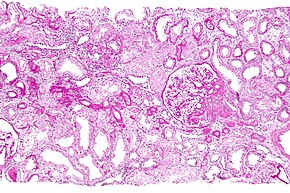
 Micrograph showing a renal core biopsy. PAS stain. | |
| ICD-9-CM | 55.23-55.24 |
| MedlinePlus | 003907 |
Renal biopsy (also kidney biopsy) is a medical procedure in which a small piece of kidney is removed from the body for examination, usually under a microscope. Microscopic examination of the tissue can provide information needed to diagnose, monitor or treat problems of the kidney.
A renal biopsy can be targeted to a particular lesion, for example a tumour arising from the kidney (targeted renal biopsy). More commonly, however, the biopsy is non-targeted as medical conditions affecting the kidney typically involve all kidney tissue indiscriminately.
A native renal biopsy is one in which the patient's own kidneys are biopsied. In a transplant renal biopsy, the kidney of another person that has been transplanted into the patient is biopsied.
Indications
Renal biopsy is recommended for selected patients with kidney disease. It is most commonly performed when less invasive measures are insufficient. The following are examples of the most common reasons for biopsy:
- Hematuria with renal dease — blood in the urine can occur with a number of conditions that affect the kidneys and urinary tract. While renal biopsy is not indicated in all cases of hematuria, it may be performed in those with hematuria as well as progressive renal disease (e.g. increasing proteinuria or blood pressure).
- Proteinuria — Proteinuria (protein in the urine) occurs in many patients with renal conditions. Renal biopsy is usually reserved for patients with relatively high or increasing levels of proteinuria or for patients who have proteinuria along with other signs of renal dysfunction.
- A patient with nephrotic syndrome (significant proteinuria, low blood albumin level, and edema (swelling) of the arms and legs) may need a renal biopsy, especially if the patient has systemic lupus erythematosus (SLE). Other patients with nephrotic syndrome may require a renal biopsy, depending upon the suspected cause of the nephrotic syndrome.
- Renal failure — impaired kidney function due to kidney injury can occur abruptly (acute renal failure) or progress over a period of time (chronic renal failure). The cause of acute renal failure can usually be determined without renal biopsy. Biopsy is performed in those instances when the cause is uncertain.
- Acute nephritic syndrome — Patients with acute nephritic syndrome have hematuria, proteinuria, high blood pressure, and impaired renal function. Renal biopsy may be recommended to determine the cause of nephritic syndrome unless it can be determined through blood testing.
Contraindications
Absolute contraindications[1]
- bleeding diathesis
- uncontrolled severe hypertension
- uncooperative patient
- presence of a solitary native kidney
Relative contraindications[2]
- azotemia
- certain anatomical abnormalities of the kidney
- skin infection at the desired biopsy site
- hemostasis-altering drugs (e.g. warfarin or heparin)
- pregnancy
- urinary tract infections
- obesity.
Procedure
Preparation — Testing may be done before the biopsy to ensure that there is no evidence of infection or a blood clotting abnormality. The biopsy is usually performed while the patient is awake, after receiving an injection of local anaesthesia (numbing medicine) to minimize pain.
To decrease the risk of bleeding, patients are usually advised to avoid medicines that increase the risk of bleeding (such as aspirin or nonsteroidal anti-inflammatory drugs (ibuprofen, naproxen)) for one to two weeks before the biopsy. If the patient takes warfarin or heparin (drugs that impair clotting and increase the risk of bleeding), the physician will give specific instructions about the dose and time to take these medications before surgery.
Biopsy procedure — In most cases, an ultrasound is done to guide the physician inserting the needle. Less commonly, computed tomography (CT scan) guidance is used. The needle is inserted through the skin in the back and into the kidney. Once the needle is in contact with the kidney, a sample of renal tissue is withdrawn.
In some patients, a different approach may be used to perform the biopsy. In this case, the patient is sedated and a small skin incision is made to obtain the sample of kidney tissue; this procedure is called open renal biopsy.
Following the biopsy, the patient made to lie flat on their back for six hours to minimize any risk of bleeding, blood pressure and urine are frequently monitored to ensure the patient is not suffering any complications.
Complications
Serious complications of renal biopsy are uncommon.
The most common complication of kidney biopsy is bleeding. This reflects the density of blood vessels within the kidney and observation that individuals with kidney failure take longer to stop bleeding after trauma (uraemic coagulopathy). Bleeding complications include a collection of blood adjacent to or around the kidney (perinephric haematoma), bleeding into the urine with passage of blood stained urine (macroscopic haematuria) or bleeding from larger blood vessels that lie adjacent the kidney. If blood clots in the bladder, this can obstruct the bladder and lead to urinary retention. The majority of bleeding that occurs following renal biopsy usually resolves on its own without long-term damage. Less commonly, the bleeding may be brisk (causing shock) or persistent (causing anaemia) or both. In these circumstances, treatment with blood transfusion or surgery may be required. Surgical options to control bleeding include less invasive catheter-delivered particles to block bleeding vessels (angioembolisation) or open surgery. In most cases, bleeding can be controlled and the kidneys are not lost. Rarely, a heavily damaged kidney may need to be removed.
Infection is rare with modern sterile operating procedures. Damage to surrounding structures, such as bowel and bladder (more likely with transplant kidney biopsy), can occur.
Occasionally, a biopsy will have to be abandoned prematurely due to technical issues such as inaccessible or small kidneys, obscured kidneys, difficult to penetrate kidneys or observation of bleeding complication. Further, after the biopsy has been completed, microscopic examination of the tissue may reveal heavily scarred tissue prompting recommendation for re-biopsy to avoid sampling error.
As with all treatments, there is a risk of allergy to the disinfectant solution, sedation, local anaesthetic and materials (latex gloves, drapes, dressings) used for the procedure.
Finally, the biopsy needle may join an artery and vein in the kidney, resulting in the formation of an arteriovenous fistula. These usually do not cause problems and close on their own. They may be monitored over time with repeat Doppler ultrasonography. Rarely, they may result in intermittent bleeding into the urine or may grow in size and threaten to burst. In these instances, the fistula may be closed surgically or with angioembolisation.
History
Until 1951, the only way of obtaining kidney tissue in a live person would be through an operation. In 1951, the Danish physicians Poul Iversen and Claus Brun described a method involving needle biopsy which has become the new standard.[3]
References
- ^ Mendelssohn D, Cole E (1995). "Outcomes of percutaneous kidney biopsy, including those of solitary native kidneys". Am J Kidney Dis. 26 (4): 580–585. PMID 7573010.
{{cite journal}}: Unknown parameter|month=ignored (help) - ^ Whittier L, Korbet S (2004). "Renal biopsy: update". Current Opinion in Nephrology and Hypertension. 13 (6): 661–665. PMID 15483458.
{{cite journal}}: Unknown parameter|month=ignored (help) - ^ Iversen P, Brun C (1951). "Aspiration biopsy of the kidney". Am. J. Med. 11 (3): 324–30. doi:10.1016/0002-9343(51)90169-6. PMID 14877837.
{{cite journal}}: Unknown parameter|month=ignored (help)
