
Human skeleton
This article includes a list of references, related reading, or external links, but its sources remain unclear because it lacks inline citations. |


The human skeleton consists of both fused and individual bones supported and supplemented by ligaments, tendons, muscles and cartilage. It serves as a scaffold which supports organs, anchors muscles, and protects organs such as the brain, lungs and heart.
The longest and heaviest bone in the body is the femur, and the smallest is the stapes bone in the middle ear. In an adult, the skeleton comprises around 20% of the total body weight.
Fused bones include those of the pelvis and the cranium. Not all bones are interconnected directly: There are 6 bones in the middle ear called the ossicles (three on each side) that articulate only with each other. The hyoid bone, which is located in the neck and serves as the point of attachment for the tongue, does not articulate with any other bones in the body, being supported by muscles and ligaments.
Development
Early in gestation, a foetus has a cartilaginous skeleton from which the long bones and most other bones gradually form throughout the remaining gestation period and for years after birth in a process called endochondral ossification. The flat bones of the skull and the clavicles are formed from connective tissue in a process known as intramembranous ossification, and ossification of the mandible occurs in the fibrous membrane covering the outer surfaces of Meckel's cartilages. At birth a newborn baby has approximately 300 bones, whereas on average an adult human has 205 bones (these numbers can vary slightly from individual to individual). The difference comes from a number of small bones that fuse together during growth, such as the sacrum and coccyx of the vertebral column. The sacrum (the bone at the base of the spine) consists of five bones which are separate at birth but fuse together into a solid structure in later years. An infant is born with zones of cartilage, called epiphyseal plates, between segments of bone to allow further growth. Growing is usually completed between ages 13 and 18, at which time the epiphyseal plates of long bones close allowing no further growth.
Segmental pattern
Much of the human skeleton maintains the ancient segmental pattern present in all vertebrates (mammals, birds, fishes, reptiles and amphibians) with basic units being repeated. This segmental pattern is particularly evident in the vertebral column and in the ribcage.
Function
The skeleton has five main functions:
Provide shape and support
The skeleton provides the framework which supports the body, and maintains its shape. The joints between bones permit movement.
Movement
Movement in vertebrates is powered by skeletal muscles, which are attached to the skeleton by tendons. Without the skeleton to give leverage, movement would be greatly restricted. However, biologically speaking, the skeleton DOES NOT enable movement.
Protection
The skeleton protects many vital organs:
- The skull protects the brain, the eyes, and the middle and inner ears.
- The spine protects the spinal cord.
- The rib cage, spine, and sternum protect the lungs, heart and major blood vessels.
- The clavicle and scapula protect the shoulder.
- The ilium and spine protect the digestive and urogenital systems and the hip.
- The patella and the ulna protect the knee and the elbow respectively.
- The carpals and tarsals protect the wrist and ankle respectively.
Blood cell production
The skeleton is the site of haematopoiesis – the generation of blood cells, which takes place in red bone marrow.
Storage
Bone matrix can store calcium and is involved in calcium metabolism, and bone marrow can store iron in ferritin and is involved in iron metabolism.
Organization
The human skeleton can be divided into the axial skeleton and the appendicular skeleton.
Gender differences

There are many differences between the male and female human skeletons. Most prominent is the difference in the pelvis, owing to characteristics required for the processes of parturition (childbirth). The shape of a female pelvis is flatter, more rounded and proportionally larger to allow the head of the fetus to pass. Men tend to have slightly thicker and longer limbs and digit bones (phalanges), while women tend to have narrower rib cages, smaller teeth, less angular mandibles, less pronounced cranial features such as the brow ridges and external occipital protuberance (the small bump at the back of the skull), and the carrying angle of the forearm is more pronounced in females.
See also
External links
Gallery
-
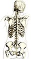 Drawing of the reverse of a female skeleton giving an impression of the location relative to surface markings
 -
 A medical drawing by a German physician from 1910
 -
 Annotated skeleton


