Cytoplasm
| Cell biology | |
|---|---|
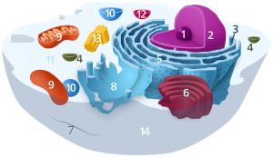
| Animal cell diagram | |
 Components of a typical animal cell:
|
The cytoplasm is the gel-like substance residing within the cell membrane holding all the cell's internal sub-structures (called organelles), outside the nucleus. All the contents of the cells of prokaryote organisms (which lack a cell nucleus) are contained within the cytoplasm. Within the cells of eukaryote organisms the contents of the cell nucleus are separated from the cytoplasm, and are then called the nucleoplasm. The cytoplasm is about 70% to 90% water and usually colourless.
It is within the cytoplasm that most cellular activities occur, such as many metabolic pathways including glycolysis, and processes such as cell division. The inner, granular mass is called the endoplasm and the outer, clear and glassy layer is called the cell cortex or the ectoplasm.
The part of the cytoplasm that is not held within organelles is called the cytosol. The cytosol is a complex mixture of cytoskeleton filaments, dissolved molecules, and water that fills much of the volume of a cell. The cytosol is a gel, with a network of fibers dispersed in water. Due to this network of fibres and high concentrations of dissolved macromolecules, such as proteins, an effect called macromolecular crowding occurs and the cytosol does not act as an ideal solution. This crowding effect alters how the components of the cytosol interact with each other.
Movement of calcium ions in and out of the cytoplasm is thought to be a signaling activity for metabolic processes.[1]
In plants, movements of the cytoplasm around vacuoles are known as cyclosis.
Constituents
The cytoplasm has three major elements; the cytosol, organelles and inclusions.
Cytosol
The cytosol is the portion of the cytoplasm not contained within membrane-bound organelles. Cytosol makes up about 70% of the cell volume and is composed of water, salts and organic molecules.[2] The cytosol also contains the protein filaments that make up the cytoskeleton, as well as soluble proteins and small structures such as ribosomes, proteasomes, and the mysterious vault complexes.[3] The inner, granular and more fluid portion of the cytoplasm is referred to as endoplasm.

Organelles
Organelles (literally "little organs"), are usually membrane-bound, and are structures inside the cell that have specific functions. Some major organelles that are suspended in the cytosol are the mitochondria, the endoplasmic reticulum, the Golgi apparatus, vacuoles, lysosomes, and in plant cells chloroplasts.
Cytoplasmic inclusions
The inclusions are small particles of insoluble substances suspended in the cytosol. A huge range of inclusions exist in different cell types, and range from crystals of calcium oxalate or silicon dioxide in plants,[4][5] to granules of energy-storage materials such as starch,[6] glycogen,[7] or polyhydroxybutyrate.[8] A particularly widespread example are lipid droplets, which are spherical droplets composed of lipids and proteins that are used in both prokaryotes and eukaryotes as a way of storing lipids such as fatty acids and sterols.[9] Lipid droplets make up much of the volume of adipocytes, which are specialized lipid-storage cells, but they are also found in a range of other cell types. cytoplasm is the only sometanse that cn sustain such force withh the nutreins and gel its made of.
Notes
- ^ C. Michael Hogan. 2010. Calcium. eds. A.Jorgensen, C. Cleveland. Encyclopedia of Earth. National Council for Science and the Environment.
- ^ Cytoplasm Composition
- ^ van Zon A, Mossink MH, Scheper RJ, Sonneveld P, Wiemer EA (2003). "The vault complex". Cell. Mol. Life Sci. 60 (9): 1828–37. doi:10.1007/s00018-003-3030-y. PMID 14523546.
{{cite journal}}: Unknown parameter|month=ignored (help)CS1 maint: multiple names: authors list (link) - ^ Prychid, Christina J.; Rudall, Paula J. (1999). "Calcium Oxalate Crystals in Monocotyledons: A Review of their Structure and Systematics". Annals of Botany. 84 (6): 725. doi:10.1006/anbo.1999.0975.
{{cite journal}}: CS1 maint: multiple names: authors list (link) - ^ Prychid, C. J.; Rudall, P. J.; Gregory, M. (2004). "Systematics and Biology of Silica Bodies in Monocotyledons". The Botanical Review. 69 (4): 377–440. doi:10.1663/0006-8101(2004)069[0377:SABOSB]2.0.CO;2.
{{cite journal}}: CS1 maint: multiple names: authors list (link) - ^ Ball SG, Morell MK (2003). "From bacterial glycogen to starch: understanding the biogenesis of the plant starch granule". Annu Rev Plant Biol. 54: 207–33. doi:10.1146/annurev.arplant.54.031902.134927. PMID 14502990.
- ^ Shearer J, Graham TE (2002). "New perspectives on the storage and organization of muscle glycogen". Can J Appl Physiol. 27 (2): 179–203. doi:10.1139/h02-012. PMID 12179957.
{{cite journal}}: Unknown parameter|month=ignored (help) - ^ Anderson AJ, Dawes EA (1 December 1990). "Occurrence, metabolism, metabolic role, and industrial uses of bacterial polyhydroxyalkanoates". Microbiol. Rev. 54 (4): 450–72. PMC 372789. PMID 2087222.
- ^ Murphy DJ (2001). "The biogenesis and functions of lipid bodies in animals, growth and microorganisms". Prog. Lipid Res. 40 (5): 325–438. doi:10.1016/S0163-7827(01)00013-3. PMID 11470496.
{{cite journal}}: Unknown parameter|month=ignored (help)
External links
- Luby-Phelps K (2000). "Cytoarchitecture and physical properties of cytoplasm: volume, viscosity, diffusion, intracellular surface area" (PDF). Int Rev Cytol. 192: 189–221.
