User:Egelberg/sandbox
Appearance
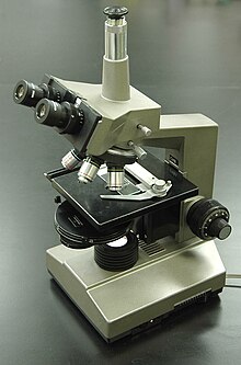
 A phase contrast microscope | |
| Uses | Microscopic observation of unstained biological material |
|---|---|
| Inventor | Frits Zernike |
| Manufacturer | Zeiss, Nikon, Olympus and others |
| Related items | Differential interference contrast microscopy, Hoffman modulation contrast microscopy, Quantitative phase contrast microscopy |
Image cytometry is a microscopy technique used by biologists to measure properties of individual cells or cell populations. Digital microscopy images are recorded and computer processed by image analysis software to count, locate or measure the morphology of cells or sub-cellular structures. Historically cells are imaged in a microscope and measured in a cytometer. An image cytometer performs these two tasks in a single device.
Cells or cellular features are often made visible through staining or by labeling with fluorescent labels.
High content screening/analysis systems, used in drug discovery to screen for drug candidates, are generally based on an image cytometer.
See also
External links
- CellProfiler — cell image analysis software by the Broad Institute


