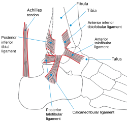
Calcaneofibular ligament
Appearance
| Calcaneofibular ligament | |
|---|---|
 The ligaments of the foot from the lateral aspect. (Label for Calcaneofibular ligament is at bottom left.) | |
 Lateral view of the human ankle | |
| Details | |
| From | calcaneus |
| To | fibula (lateral malleolus) |
| Identifiers | |
| Latin | ligamentum calcaneofibulare |
| TA98 | A03.6.10.011 |
| TA2 | 1921 |
| FMA | 44089 |
| Anatomical terminology | |
The calcaneofibular ligament is a narrow, rounded cord, running from the tip of the lateral malleolus of the fibula downward and slightly backward to a tubercle on the lateral surface of the calcaneus. It is part of the lateral collateral ligament, which opposes the hyperinversion of the subtalar joint, as in a common type of ankle sprain.[1]
It is covered by the tendons of the fibularis longus and brevis muscles.
References
![]() This article incorporates text in the public domain from page 351 of the 20th edition of Gray's Anatomy (1918)
This article incorporates text in the public domain from page 351 of the 20th edition of Gray's Anatomy (1918)
- ^ Moore KL, Dalley AF, Agur AM (2013). Clinically Oriented Anatomy (7th ed.). Lippincott Williams & Wilkins. ISBN 978-1-4511-8447-1.
Further reading
- Matsui K, Takao M, Tochigi Y, Ozeki S, Glazebrook M (June 2017). "Anatomy of anterior talofibular ligament and calcaneofibular ligament for minimally invasive surgery: a systematic review". Knee Surgery, Sports Traumatology, Arthroscopy (Review). 25 (6): 1892–1902. doi:10.1007/s00167-016-4194-y. PMID 27295109.
External links
- Calcaneofibular ligament at the Duke University Health System's Orthopedics program
- sports/14 at eMedicine—Calcaneofibular ligament injury
- lljoints at The Anatomy Lesson by Wesley Norman (Georgetown University) (posterioranklejoint)
- Anatomy figure: 17:10-05 at Human Anatomy Online, SUNY Downstate Medical Center
