Alar fascia
Appearance
| Alar fascia | |
|---|---|
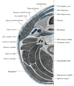
 Section of the neck at about the level of the sixth cervical vertebra. Showing the arrangement of the fascia coli. | |
| Details | |
| Identifiers | |
| Latin | lamina alaris fasciae cervicalis |
| TA2 | 2217 |
| Anatomical terminology | |
The alar fascia is a layer of fascia, sometimes described as part of the prevertebral fascia,[1] and sometimes as in front of it.
Cranially, it reaches the skull, and caudally, it reaches the second thoracic vertebra.[2]
It is the posterior border of the retropharyngeal space.[3]
In 2015, the anatomy of the alar fascia was revisited using dissection in conjunction with E12 plastination. The authors revealed that the alar fascia originated as a well defined midline structure at the level of C1 and does not reach the base of the skull. It is suggested that the area between C1 and the base of the skull is a potential entry into the danger space.[4]
See also
References
- ^ "Untitled Document". Archived from the original on 2008-02-14. Retrieved 2008-02-19.
- ^ Kyung Won, PhD. Chung (2005). Gross Anatomy (Board Review). Hagerstown, MD: Lippincott Williams & Wilkins. p. 362. ISBN 0-7817-5309-0.
- ^ Ozlugedik S, Ibrahim Acar H, Apaydin N, et al. (October 2005). "Retropharyngeal space and lymph nodes: an anatomical guide for surgical dissection". Acta Otolaryngol. 125 (10): 1111–5. doi:10.1080/00016480510035421. PMID 16298795.
- ^ Frank Scali; Lance G Nash; Matthew E Pontell (2015). "Defining the Morphology and Distribution of the Alar Fascia: A Sheet Plastination Investigation". Annals of Otology, Rhinology, and Laryngology. 124: 814–9. doi:10.1177/0003489415588129. PMID 25991834.
