Accessory hemiazygos vein
| Accessory hemiazygos vein | |
|---|---|
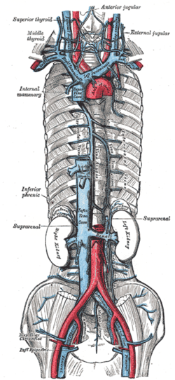
 Diagram showing completion of development of the parietal veins (accessory hemiazygos vein visible at center-left) | |
 The venae cavae and azygos veins, with their tributaries. | |
| Details | |
| Drains to | Hemiazygos vein, azygos vein |
| Identifiers | |
| Latin | Vena hemiazygos accessoria, vena azygos minor superior |
| TA98 | A12.3.07.005 |
| TA2 | 4758 |
| FMA | 5011 |
| Anatomical terminology | |
The accessory hemiazygos vein, also called the superior hemiazygous vein[1]is a vein on the left side of the vertebral column that generally drains the fourth through eighth intercostal spaces on the left side of the body.[2]
Structure
The accessory hemiazygos vein varies inversely in size with the left superior intercostal vein.
It receives the posterior intercostal veins from the 4th, 5th, 6th, and 7th intercostal spaces between the left superior intercostal vein and highest tributary of the hemiazygos vein; the left bronchial vein sometimes opens into it.
It either crosses the body of the eighth thoracic vertebra to join the azygos vein or ends in the hemiazygos.
When this vein is small, or altogether absent, the left superior intercostal vein may extend as low as the fifth or sixth intercostal space.
References
- ^ Blackmon JM, Franco A (July 2011). "Normal variants of the accessory hemiazygos vein". The British Journal of Radiology. 84 (1003): 659–60. doi:10.1259/bjr/13695502. PMC 3473485. PMID 21697414.
- ^ Dahran N, Soames R (September 2016). "Anatomical Variations of the Azygos Venous System: Classification and Clinical Relevance". International Journal of Morphology. 34 (3): 1128–36. doi:10.4067/S0717-95022016000300051.
