Ameloblast
| Ameloblast | |
|---|---|
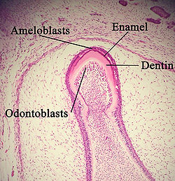
 A developing tooth with odontoblasts marked. | |
 The cervical loop area: (1) dental follicle cells, (2) dental mesenchyme, (3) Odontoblasts, (4) Dentin, (5) stellate reticulum, (6) outer enamel epithelium, (7)inner enamel epithelium, (8) ameloblasts, (9) enamel. | |
| Details | |
| Identifiers | |
| Latin | ameloblastus |
| MeSH | D000565 |
| TE | E5.4.1.1.2.3.20 |
| FMA | 70576 |
| Anatomical terminology | |
Ameloblasts are cells, present only during tooth development, that deposit tooth enamel, the hard outermost layer of the tooth that forms the chewing surface.
Ameloblasts are cells which secrete the enamel proteins enamelin and amelogenin which will later mineralize to form enamel on teeth, the hardest substance in the human body.[1] Each Ameloblast is approximately 4 micrometers in diameter, 40 micrometers in length and has a hexagonal cross section. The secretory end of the ameloblast ends in a six-sided pyramid-like projection known as the Tomes' process. The angulation of the Tomes' process is significant in the orientation of enamel rods. Ameloblasts are derived from oral epithelium tissue of ectodermal origin. Their differentiation from preameloblasts is a result of signaling from the ectomesenchymal cells of the dental papilla. The ameloblasts will only become fully functional after the first layer of dentine has been formed by odontoblasts. Ameloblasts control ionic and organic compositions of enamel. They adjust their secretory and resorptive activities to maintain favorable conditions for biomineralization.
The murine ALC cell line is of ameloblastic origin.[2]
Life cycle of ameloblasts
The life cycle of ameloblasts consists of six stages :
- Morphogenic stage
- Organizing stage
- Formative (secretory) stage (Tome's processes appear in secretory stage)
- Maturative stage
- Protective stage
- Desmolytic stage
References
- ^ Gartner, Leslie P., James L. Hiatt. (2001). Color Textbook of Histology, 2nd Edition. Philadelphia: W.B. Saunders Company. p. 366. ISBN 0-7216-8806-3.
{{cite book}}: CS1 maint: multiple names: authors list (link) - ^ Takahashi, Satomi; Kawashima, Nobuyuki; Sakamoto, Kei; Nakata, Akira; Kameda, Takashi; Sugiyama, Toshihiro; Katsube, Ken-Ichi; Suda, Hideaki (2007). "Differentiation of an ameloblast-lineage cell line (ALC) is induced by Sonic hedgehog signaling". Biochemical and Biophysical Research Communications. 353 (2): 405–11. doi:10.1016/j.bbrc.2006.12.053. PMID 17188245.
See also
Template:Human cell types derived primarily from ectoderm
