Capsule endoscopy
| Wireless Capsule Endoscopy | |
|---|---|
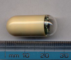
 Picture of a capsule | |
| MeSH | D053704 |
| OPS-301 code | 1-63a, 1-656 |


Capsule endoscopy is a way to record images of the digestive tract for use in medicine. The capsule is the size and shape of a pill and contains a tiny camera. After a patient swallows the capsule, it takes pictures of the inside of the gastrointestinal tract. The primary use of capsule endoscopy is to examine areas of the small intestine that cannot be seen by other types of endoscopy such as colonoscopy or esophagogastroduodenoscopy (EGD).
Medical uses
Capsule endoscopy is used to examine parts of the gastrointestinal tract that cannot be seen with other types of endoscopy. Upper endoscopy, also called EGD, uses a camera attached to a long flexible tube to view the esophagus, the stomach and the beginning of the first part of the small intestine called the duodenum. A colonoscope, inserted through the rectum,can view the colon and the distal portion of the small intestine, the terminal ileum. These two types of endoscopy cannot visualize the majority of the middle portion of the gastrointestinal tract, the small intestine. Capsule endoscopy is useful when disease is suspected in the small intestine, and can sometimes diagnose sources of occult bleeding [blood visible microscopically only] or causes of abdominal pain such as Crohn's disease, or peptic ulcers. Capsule endoscopy can be used to diagnose problems in the small intestine, but unlike EGD or colonoscopy it cannot treat pathology that may be discovered. Capsule endoscopy transfers the captured images wirelessly to an external receiver worn by the patient using one of appropriate frequency bands. The collected images are then transferred to a computer for diagnosis, review and display.[1] A transmitted radio-frequency signal can be used to accurately estimate the location of the capsule and to track it in real time inside the body and gastrointestinal tract.[2]
Common reasons for doing Capsule Endoscopy include unexplained bleeding, unexplained iron deficiency, unexplained abdominal pain, search for polyps, ulcers and tumors of small intestine and inflammatory bowel disease such as Crohn's disease.[3]
Current attempts in the area of wireless capsule endoscopy are targeting to include additional sensing mechanisms, localization,and motion control enabling new applications, e.g drug delivery. Wireless energy transmission is also investigated for wireless capsule systems to provide a continuous energy source.[4]
It is unclear if capsule endoscopy can replace gastroscopy in those with cirrhosis.[5]
Side effects
It is considered to be a very safe method to determine an unknown cause of a gastrointestinal bleed.[6] The capsule usually passes through feces within 24–48 hours.[6] There has been a report of retention of the capsule for almost 5 years. The patient was asymptomatic.[6]
References
- ^ M. R. Yuce and T. Dissanayake (2012). "Easy-to-swallow wireless telemetry". IEEE Microwave Magazine. Retrieved 18 May 2015.
- ^ M. Pourhomayoun, M. L. Fowler and Z. Jin (2012). "A Novel Method for Medical Implant In-Body Localization" (PDF). Proceedings of the IEEE International Conference of Engineering in Medicine and Biology. Retrieved 30 December 2012.
- ^ http://www.laintegrativegi.com/technical-services.html#Capsule-Endoscopy
- ^ M. R. Yuce, G. Alici and T.D. Than (2014). "Wireless Endoscopy". Wiley Encyclopedia of Electrical and Electronics Engineering. Retrieved December 2014.
{{cite journal}}: Check date values in:|accessdate=(help) - ^ Colli, A; Gana, JC; Turner, D; Yap, J; Adams-Webber, T; Ling, SC; Casazza, G (Oct 1, 2014). "Capsule endoscopy for the diagnosis of oesophageal varices in people with chronic liver disease or portal vein thrombosis". The Cochrane database of systematic reviews. 10: CD008760. doi:10.1002/14651858.CD008760.pub2. PMID 25271409.
- ^ a b c http://www.ncbi.nlm.nih.gov/pmc/articles/PMC3711067/
