Jump to content

File:Hemoglobin F.png

Page contents not supported in other languages.
This is a file from the Wikimedia Commons
From Wikipedia, the free encyclopedia

Hemoglobin_F.png (600 × 600 pixels, file size: 294 KB, MIME type: image/png)

Summary

Description
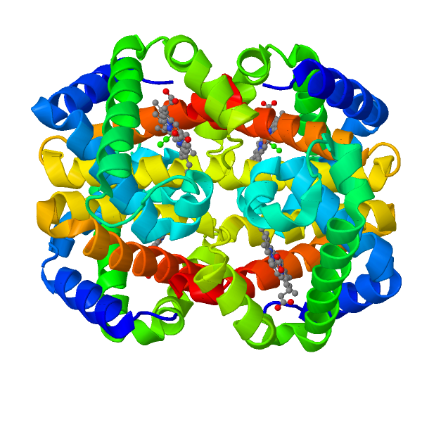
English: Fetal hemoglobin (HbF), formed by two alpha subunits (top) and two gamma subunits (bottom). The 4 heme groups are also displayed. All chains (ribbons) are rainbow-colored from blue to red (N- to C-termini)
Date 10 February 2016 (original upload date)
Source Own work - A rendering prepared by me, using Jmol, from 4MQJ at PDB (doi:10.2210/pdb4mqj/pdb).
Author AngelHerraez

Licensing

Public domain I, the copyright holder of this work, release this work into the public domain. This applies worldwide.
In some countries this may not be legally possible; if so:
I grant anyone the right to use this work for any purpose, without any conditions, unless such conditions are required by law.

Original upload log

The original description page was here. All following user names refer to en.wikipedia.
  • 2004-04-12 15:39 Diberri 612×522× (25345 bytes) Fetal hemoglobin (transparent background). My snapshot of rendered version from PDB file [http://www.snv.jussieu.fr/vie/telecharge/pdb/HbF-1FDH.pdb].
  • 2004-04-12 15:01 Diberri 612×522× (36662 bytes) Fetal hemoglobin (white background). My snapshot of rendered version from PDB file [http://www.snv.jussieu.fr/vie/telecharge/pdb/HbF-1FDH.pdb].

Captions

Fetal hemoglobin (HbF), formed by two alpha subunits (top) and two gamma subunits (bottom). The 4 heme groups are also displayed. All chains (ribbons) are rainbow-colored from blue to red (N- to C-termini)

Items portrayed in this file

depicts

10 February 2016

301,544 byte

600 pixel

600 pixel

image/png

1ba491b9a55606ad20d91578cecea55292e76022

File history

Click on a date/time to view the file as it appeared at that time.

Date/TimeThumbnailDimensionsUserComment
current12:25, 10 February 2016Thumbnail for version as of 12:25, 10 February 2016600 × 600 (294 KB)AngelHerraezthe former image was partially clipped
12:21, 10 February 2016Thumbnail for version as of 12:21, 10 February 2016600 × 600 (360 KB)AngelHerraezThe prevoius image was a dimer (alpha, gamma) and hence may be misleading. The actual, biologically relevant, structure of hemoglobin F (fetal) is a tetramer (2 alpha + 2 gamma subunits). This rendering displays the tetramer, as well as the 4 heme grou...
07:06, 16 January 2012Thumbnail for version as of 07:06, 16 January 2012612 × 522 (25 KB)File Upload Bot (Magnus Manske) {{BotMoveToCommons|en.wikipedia|year={{subst:CURRENTYEAR}}|month={{subst:CURRENTMONTHNAME}}|day={{subst:CURRENTDAY}}}} {{Information |Description={{en|Fetal hemoglobin (white background).}} |Source=Transferred from [http://en.wikipedia.org en.wikipedia];

Global file usage

The following other wikis use this file:

Metadata