
Fragment antigen-binding region

The antigen-binding fragment (Fab) is a region on an antibody that binds to antigens. It is composed of one constant and one variable domain of each of the heavy and the light chain. The variable domain contains the paratope (the antigen-binding site), comprising a set of complementarity determining regions, at the amino terminal end of the monomer. Each arm of the Y thus binds an epitope on the antigen.
Preparation
In an experimental setting, Fc and Fab fragments can be generated in the laboratory. The enzyme papain can be used to cleave an immunoglobulin monomer into two Fab fragments and an Fc fragment. The enzyme pepsin cleaves below the hinge region, so a F(ab')2 fragment and a pFc' fragment is formed. Recently another enzyme for generation of F(ab')2 has been commercially available. The enzyme IdeS (Immunoglobulin degrading enzyme from Streptococcus pyogenes, trade name FabRICATOR) cleaves IgG in a sequence specific manner at neutral pH. The F(ab')2 fragment can be split into two Fab' fragments by mild reduction.[1]
-
 Heavy and light chains, variable and constant regions of an antibody.
 -
 An antibody digested by papain yields three fragments: two Fab fragments and one Fc fragment
 -
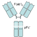 An antibody digested by pepsin yields two fragments: a F(ab')2 fragment and a pFc' fragment


The variable regions of the heavy and light chains can be fused together to form a single-chain variable fragment (scFv), which is only half the size of the Fab fragment, yet retains the original specificity of the parent immunoglobulin.[2]
Applications
Fabs have seen some theraputic use in emergency medicine as an antidote. Marketed applications include Digoxin immune fab and Crofab, a mixture of Fabs for rattlesnake bites. Fabs against colchicine and tricyclic antidepressants has also been produced but are yet to see approval.[3][4]
Fabs are a common form-factor for monoclonal antibodies designated for theraputic use. The Fab abciximab, which inhibits blood clotting, works by disabling Glycoprotein IIb/IIIa fount on platelets.[5] Ranibizumab, a treatment for macular degeneration, targets vascular endothelial growth factor A, a protein involved in the growth of blood vessels. Certolizumab pegol is a Fab chemically linked to PEG, and it treats various inflammatory disorders by binding away TNFα.
Fab antibodies also have diagnostic use. Arcitumomab is a mouse antibody that recognizes Carcinoembryonic antigen, an antigen over-expressed in 95% of colorectal cancers. It is conjugated to a radioactive element, which will label the tumors when viewed with single photon emission computed tomography. Sulesomab, an antigen that recognizes proteins on the surface of granulocytes, is used to label out infections, again using the 99mTc isotope.[6]
See also
References
- ^ Larsson, Lars-Inge (September 1988). Immunocytochemistry: Theory and practice. Crc Press. p. 1. ISBN 0-8493-6078-1.
- ^ Janeway, CA, Jr.; et al. (2001). Immunobiology (5th ed.). Garland Publishing. ISBN 0-8153-3642-X.
{{cite book}}: CS1 maint: multiple names: authors list (link) - ^ Flanagan RJ, Jones AL (2004). "Fab antibody fragments: some applications in clinical toxicology". Drug Saf. 27 (14): 1115–1133. PMID 15554746.
- ^ Seger D, Kahn S, Krenzelok EP (2005). "Treatment of US crotalidae bites: comparisons of serum and globulin-based polyvalent and antigen-binding fragment antivenins". Toxicol Rev. 24 (4): 217–227. PMID 16499404.
{{cite journal}}: CS1 maint: multiple names: authors list (link) - ^ Fachinformation Lucentis®. Novartis Pharma. Stand 15. November 2007.
- ^ W. J. Köstler, C. C. Zielinski (November 2000). "Diagnostische und therapeutische Antikörper in der Onkologie — State of the Art". Acta Chirurgica Austriaca. 32 (6): 260–263.
Engineered monoclonal antibodies and antibody mimetics | ||
|---|---|---|
| Whole antibody |  | |
| Fab fragment | ||
| Variable fragment | ||
| Smaller units | ||
| Intracellular | ||
| Antibody mimetics | ||
This immunology article is a stub. You can help Wikipedia by expanding it. |