
Squamous-cell carcinoma
Squamous cell carcinomas, also known as epidermoid carcinoma are a number of different types of cancer that result from squamous cells.[1] These cells form the surface of the skin lining of hollow organs in the body and line the respiratory and digestive tracts.[1]
Common types include:
- Squamous cell skin cancer: A type of skin cancer
- Squamous-cell carcinoma of the lung: A type of lung cancer
- Squamous cell thyroid carcinoma: A type of thyroid cancer
- Esophageal squamous cell carcinoma: A type of esophageal cancer
Despite sharing the name squamous cell carcinoma, the SCCs of different body sites can show differences in their presented symptoms, natural history, prognosis, and response to treatment.
Types
Human papillomavirus infection (HPV) has been associated with SCC of the oropharynx, lung,[2] fingers[3] and anogenital region.
Head and neck cancer

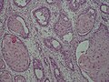
_squamous_cell_carcinoma_histopathology.jpg)
Ninety percent[4] of cases of head and neck cancer (cancer of the mouth, nasal cavity, nasopharynx, throat and associated structures) are due to squamous cell carcinoma.
Thyroid
Primary squamous cell thyroid carcinoma shows an aggressive biological phenotype resulting in poor prognosis for patients.[5]
Esophagus
Esophageal cancer may be due to either squamous cell carcinoma (ESCC) or adenocarcinoma (EAC). SCCs tend to occur closer to the mouth, while adenocarcinomas occur closer to the stomach. Dysphagia (difficulty swallowing, solids worse than liquids) and painful swallowing are common initial symptoms. If the disease is localized, surgical removal of the affected esophagus may offer the possibility of a cure. If the disease has spread, chemotherapy and radiotherapy are commonly used.
Lung

When associated with the lung, it is typically a centrally located large cell cancer (non-small cell lung cancer or NSCLC). It often has a paraneoplastic syndrome causing ectopic production of parathyroid hormone-related protein (PTHrP), resulting in hypercalcemia, however paraneoplastic syndrome is more commonly associated with small cell lung cancer.
It is primarily due to smoking.[6]
Penis
Human papillomavirus (HPV), primarily HPV 16 & 18, are strongly implicated in the development of squamous cell carcinoma of the penis.
Three carcinoma in situ are associated with squamous cell carcinoma of the penis:
1) Bowen's disease which presents as leukoplakia on the shaft. Around 1/3 progress to squamous cell carcinoma
2) Erythroplasia of Queyrat, a variation of Bowen's Disease, presenting as erythroplakia on the glans
3) Bowenoid papulosis, which histologically resembles Bowen disease, but presents as reddish papules.[7]
Prostate
When associated with the prostate, squamous cell carcinoma is very aggressive in nature. It is difficult to detect as there is no increase in prostate specific antigen levels seen; meaning that the cancer is often diagnosed at an advanced stage.
Vagina and cervix
Vaginal squamous cell carcinoma spreads slowly and usually stays near the vagina, but may spread to the lungs and liver. This is the most common type of vaginal cancer.
Bladder
Most bladder cancer is transitional cell, but bladder cancer associated with Schistosomiasis is often squamous cell carcinoma.
Classification
Cancer can be considered a very large and exceptionally heterogeneous family of malignant diseases, with squamous cell carcinomas comprising one of the largest subsets.[8][9][10] All squamous cell carcinoma lesions are thought to begin via the repeated, uncontrolled division of cancer stem cells of epithelial lineage or characteristics.[citation needed] Squamous cell carcinomas arise from squamous cells, which are flat cells that line many areas of the body. Accumulation of these cancer cells causes a microscopic focus of abnormal cells that are, at least initially, locally confined within the specific tissue in which the progenitor cell resided. This condition is called squamous cell carcinoma in situ, and it is diagnosed when the tumor has not yet penetrated the basement membrane or other delimiting structure to invade adjacent tissues. Once the lesion has grown and progressed to the point where it has breached, penetrated, and infiltrated adjacent structures, it is referred to as "invasive" squamous cell carcinoma. Once a carcinoma becomes invasive, it is able to spread to other organs and cause the formation of a metastasis, or "secondary tumor".
Tissue of origin
The International Classification of Diseases for Oncology (ICD-O) system lists a number of morphological subtypes and variants of malignant squamous cell neoplasms, including:[11]
- Papillary thyroid carcinoma (Code 8050/3)
- Verrucous squamous cell carcinoma (Code 8051/3)
- Papillary squamous cell carcinoma (Code 8052/3)
- Squamous cell carcinoma (Code 8070/3)
- Large cell keratinizing squamous cell carcinoma (Code 8071/3)
- Large cell nonkeratinizing squamous cell carcinoma (Code 8072/3)
- Small cell keratinizing squamous cell carcinoma (Code 8073/3)
- Spindle cell squamous cell carcinoma(Code 8074/3)
- Adenoid/pseudoglandular squamous cell carcinoma(Code 8075/3)
- Intraepidermal squamous cell carcinoma (Code 8081/3)
- Lymphoepithelial carcinoma (Code 8082/3)
Other variants of squamous cell carcinoma are recognized under other systems, such as:
Morphology
- Bowen's disease is a sunlight-induced skin disease, and is considered to be an early form of squamous cell carcinoma.
- Erythroplasia of Queyrat
- Keratoacanthoma is a low-grade malignancy of the skin. It originates in the pilo-sebaceous glands, and is similar in clinical presentation and microscopic analysis to squamous cell carcinoma, except that it contains a central keratin plug. Statistically, it is less likely to become invasive than squamous cell carcinoma.
- Marjolin's ulcer is a type of squamous cell carcinoma that arises from a non-healing ulcer or burn wound. More recent evidence, however, suggests that there may be genetic differences between squamous cell carcinoma and marjolin's ulcer which were previously underappreciated.[12]
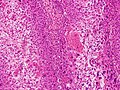
Microscopic appearance
One method of classifying squamous cell carcinomas is by their appearance under microscope. Subtypes may include:
- Adenoid squamous cell carcinoma' (also known as "Pseudoglandular squamous cell carcinoma"), characterized by a tubular microscopic pattern and keratinocyte acantholysis.[13]
- Basaloid squamous cell carcinoma is characterized by a predilection for the tongue base.[13]
- Clear-cell squamous cell carcinoma (also known as "Clear-cell carcinoma of the skin") is characterized by keratinocytes that appear clear as a result of hydropic swelling.[13]
- Signet-ring-cell squamous cell carcinoma (occasionally rendered as "signet-ring-cell squamous cell carcinoma") is a histological variant characterized by concentric rings composed of keratin and large vacuoles corresponding to markedly dilated endoplasmic reticulum.[13] These vacuoles grow to such an extent that they radically displace the cell nucleus toward the cell membrane, giving the cell a distinctive superficial resemblance to a "signet ring" when viewed under a microscope.
- Spindle-cell squamous cell carcinoma (also known as "Spindle-cell carcinoma"[14]) is a subtype characterized by spindle-shaped atypical cells.[13]
SCC is a histologically distinct form of cancer. It arises from the uncontrolled multiplication of cells of epithelium, or cells showing particular cytological or tissue architectural characteristics of squamous cell differentiation, such as the presence of keratin, tonofilament bundles, or desmosomes, structures involved in cell-to-cell adhesion.
-
 Adenoid squamous cell carcinoma
 -
 Basaloid squamous cell carcinoma
 -
 Clear-cell squamous cell carcinoma
 -
 Spindle-cell squamous cell carcinoma


References
- ^ a b "NCI Dictionary of Cancer Terms". National Cancer Institute. Retrieved 9 November 2016.
- ^ Yu Y, Yang A, Hu S, Yan H (June 2009). "Correlation of HPV-16/18 infection of human papillomavirus with lung squamous cell carcinomas in Western China". Oncol. Rep. 21 (6): 1627–32. doi:10.3892/or_00000397. PMID 19424646.
- ^ "Recurrent Squamous Cell Carcinoma In Situ of the Finger". Retrieved 2010-09-22.
- ^ "Types of head and neck cancer - Understanding - Macmillan Cancer Support". Retrieved 15 March 2017.
- ^ MI Syed; M Stewart; S Syed; S Dahill; C Adams; DR Mclellan; LJ Clark. (2011). "Squamous cell carcinoma of the thyroid gland: primary or secondary disease?". The Journal of Laryngology & Otology. 125 (1): 3–9. doi:10.1017/S0022215110002070. PMID 20950510.
- ^ al.], [edited by] Ruth A. Hannon ... [et (2010). Porth pathophysiology : concepts of altered health states (1st Canadian ed.). Philadelphia, PA: Wolters Kluwer Health/Lippincott Williams & Wilkins. p. 660. ISBN 978-1-60547-781-7.
{{cite book}}:|first=has generic name (help) - ^ Robbins, Stanley; Kumar, Vinay; Abbas, Abul; Fausto, Nelson (2007). Robbins Basic Pathology (8th ed. ed.). Philadelphia: Saunders/Elsevier. p. 688. ISBN 978-1-4160-2973-1.
{{cite book}}:|edition=has extra text (help) - ^ Berman JJ (November 2004). "Tumor taxonomy for the developmental lineage classification of neoplasms". BMC Cancer. 4 (1): 88. doi:10.1186/1471-2407-4-88. PMC 535937. PMID 15571625.
{{cite journal}}: CS1 maint: unflagged free DOI (link) - ^ Berman JJ (March 2004). "Tumor classification: molecular analysis meets Aristotle". BMC Cancer. 4 (1): 10. doi:10.1186/1471-2407-4-10. PMC 415552. PMID 15113444.
{{cite journal}}: CS1 maint: unflagged free DOI (link) - ^ Travis, William D; Brambilla, Elisabeth; Muller-Hermelink, H Konrad; et al., eds. (2004). Pathology and Genetics of Tumours of the Lung, Pleura, Thymus and Heart (PDF). World Health Organization Classification of Tumours. Lyon: IARC Press. ISBN 92-832-2418-3. Retrieved 27 March 2010.
- ^ World Health Organization. International Classification of Diseases for Oncology, Second Edition. Geneva, Switzerland: World Health Organization, 1990.
- ^ Sinha S, Su S, Workentine M, Agabalyan N, Cheng M, Gabriel V, Biernaskie J (January 2017). "Transcriptional Analysis Reveals Evidence of Chronically Impeded ECM Turnover and Epithelium-to-Mesenchyme Transition in Scar Tissue Giving Rise to Marjolin's Ulcer". J Burn Care Res. 38 (1): e14–e22. doi:10.1097/BCR.0000000000000432. PMID 27679957.
- ^ a b c d e Freedberg, Irwin M., ed. (2003). Fitzpatrick's dermatology in general medicine (6th ed.). New York, NY [u.a.]: McGraw-Hill. p. 743. ISBN 0-07-138076-0.
{{cite book}}: Unknown parameter|displayeditors=ignored (|display-editors=suggested) (help) - ^ Rapini, Ronald P.; Bolognia, Jean L.; Jorizzo, Joseph L. (2007). Dermatology: 2-Volume Set. St. Louis: Mosby. ISBN 1-4160-2999-0.