Thoracic splanchnic nerves
Appearance
| Thoracic splanchnic nerves | |
|---|---|
 The right sympathetic chain and its connections with the thoracic, abdominal, and pelvic plexuses. (Greater and lesser splanchnic nerves labeled at left.) | |
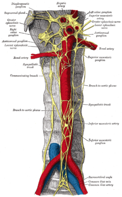
 Abdominal portion of the sympathetic trunk, with the celiac and hypogastric plexuses. (Greater splanchnic and lowest splanchnic labeled at upper left. Greater splanchnic and lesser splanchnic labeled at upper right.) | |
| Details | |
| From | thoracic ganglia |
| Identifiers | |
| TA98 | A14.3.01.028 A14.3.01.032 A14.3.01.030 |
| TA2 | 6631, 6632, 6634 |
| FMA | 6280 |
| Anatomical terms of neuroanatomy | |
Thoracic splanchnic nerves are splanchnic nerves that arise from the sympathetic trunk in the thorax and travel inferiorly to provide sympathetic innervation to the abdomen. The nerves contain preganglionic sympathetic and general visceral afferent fibers.
There are three main thoracic splanchnic nerves:
| Name | Ganglia | Description |
|---|---|---|
| greater[1] | T5-T9 or T5-T10[2] | The nerve travels through the diaphragm and enters the abdominal cavity, where its fibers synapse at the celiac ganglia. The nerve contributes to the celiac plexus, a network of nerves located in the vicinity of where the celiac trunk branches from the abdominal aorta. The fibers in this nerve modulate the activity of the enteric nervous system of the foregut. They also provide the sympathetic innervation to the adrenal medulla, stimulating catecholamine release. |
| lesser[3] | T9-T12, T9-T10,[3] T10-T12, or T10-T11[2] | The nerve travels inferiorly, lateral to the greater splanchnic nerve. Its fibers synapse with their postganglionic counterparts in the superior mesenteric ganglia, or in the aorticorenal ganglion. The nerve modulates the activity of the enteric nervous system of the midgut. |
| least or lowest[4] | T12-L2, or T11-T12 | The nerve travels into the abdomen, where its fibers synapse in the renal ganglia. |
The nerve's origins can be remembered by the "4-3-2 rule", accounting for the number of ganglia giving rise to each nerve. However, different sources define the nerves in different ways, so this rule may not always be reliable.
Additional images
-
Greater splanchnic nerve, seen in thoracic cavity seen from left side.
-
Diagram of efferent sympathetic nervous system.
-
Plan of right sympathetic cord and splanchnic nerves.
-
The celiac ganglia with the sympathetic plexuses of the abdominal viscera radiating from the ganglia.
-
Lower half of right sympathetic cord.
-
The relations of the viscera and large vessels of the abdomen. Seen from behind, the last thoracic vertebra being well raised.
-
Thoracic splanchnic nerves
References
- ^ "greater splanchnic nerve" at Dorland's Medical Dictionary
- ^ a b thoraxlesson5 at The Anatomy Lesson by Wesley Norman (Georgetown University)
- ^ a b "lesser splanchnic nerve" at Dorland's Medical Dictionary
- ^ "Least splanchnic nerve" at Dorland's Medical Dictionary
External links
- Anatomy figure: 21:04-07 at Human Anatomy Online, SUNY Downstate Medical Center - "The position of the right and left vagus nerves, and sympathetic trunks in the mediastinum."
- Anatomy photo:40:10-0102 at the SUNY Downstate Medical Center - "Posterior Abdominal Wall: The Celiac Plexus"
- . GPnotebook https://www.gpnotebook.co.uk/simplepage.cfm?ID=-1274675140.
{{cite web}}: Missing or empty|title=(help) - figures/chapter_30/30-4.HTM: Basic Human Anatomy at Dartmouth Medical School
- figures/chapter_32/32-6.HTM: Basic Human Anatomy at Dartmouth Medical School


