Thanatotranscriptome: Difference between revisions
Citation bot (talk | contribs) Alter: pmc. Add: s2cid, pmid, authors 1-1. Removed proxy/dead URL that duplicated identifier. Removed parameters. Some additions/deletions were parameter name changes. | Use this bot. Report bugs. | Suggested by RoanokeVirginia | #UCB_toolbar |
mNo edit summary |
||
| Line 10: | Line 10: | ||
It can from a [[serology]] postmortem characterize [[transcriptome]] of [[tissue (biology)|tissue]] particular cell type, or compare the transcriptomes between various conditions experimental. |
It can from a [[serology]] postmortem characterize [[transcriptome]] of [[tissue (biology)|tissue]] particular cell type, or compare the transcriptomes between various conditions experimental. |
||
It can be complementary to the analysis of thanatomicrobiome to better understand the process of transformation of the necromass in the days following the death.<ref>Javan |
It can be complementary to the analysis of thanatomicrobiome to better understand the process of transformation of the necromass in the days following the death.<ref>{{Cite journal |last=Javan |first=Gulnaz T. |last2=Finley |first2=Sheree J. |last3=Abidin |first3=Zain |last4=Mulle |first4=Jennifer G. |date=2016-02-24 |title=The Thanatomicrobiome: A Missing Piece of the Microbial Puzzle of Death |url=https://www.frontiersin.org/article/10.3389/fmicb.2016.00225 |journal=Frontiers in Microbiology |volume=7 |doi=10.3389/fmicb.2016.00225 |issn=1664-302X |pmc=PMC4764706 |pmid=26941736}}</ref> |
||
'''The human thanatomicrobiome''' is an original term introduced '''by Dr. Gulnaz Javan in 2014''' at the 66th Annual Meeting of the American Academy of Forensic Sciences in Seattle, Washington.{{Citation needed}} The thanatomicrobiome (''thanatos''—Greek for death) is characterized by a diverse assortment of microorganisms located in internal organs (brain, heart, liver, and spleen) and blood samples collected after a human dies. It is defined as the microbial community of internal body sites, created by a successional process whereby trillions of microorganisms populate, proliferate, and/or die within the dead body, resulting in temporal modifications in the community composition over time. |
'''The human thanatomicrobiome''' is an original term introduced '''by Dr. Gulnaz Javan in 2014''' at the 66th Annual Meeting of the American Academy of Forensic Sciences in Seattle, Washington.{{Citation needed}} The thanatomicrobiome (''thanatos''—Greek for death) is characterized by a diverse assortment of microorganisms located in internal organs (brain, heart, liver, and spleen) and blood samples collected after a human dies. It is defined as the microbial community of internal body sites, created by a successional process whereby trillions of microorganisms populate, proliferate, and/or die within the dead body, resulting in temporal modifications in the community composition over time. |
||
| Line 27: | Line 27: | ||
This information could possibly in the future lead to: |
This information could possibly in the future lead to: |
||
* Construct a definition both more accurate and nuanced phenomenon of "[[death]]" |
* Construct a definition both more accurate and nuanced phenomenon of "[[death]]". |
||
* More precise time of death by the [[forensic]] (or a biologist or veterinarian in an [[EcoHealth]] investigation who needs information on hours or causes of poisoning, without the case of a [[zoonosis]] for example. <br />We are far, but if we come to better understand the steps of this phenomenon in the [[Human]], a coroner could via a ''"postmortem serology"''<ref>Moreno, L. I., Tate, C. M., Knott, E. L., McDaniel, J. E., Rogers, S. S., Koons, B. W., ... & Robertson, J. M. (2012). Determination of an effective housekeeping gene for the quantification of mRNA for forensic applications. Journal of Forensic Sciences, 57(4), 1051-1058 ([http://onlinelibrary.wiley.com/doi/10.1111/j.1556-4029.2012.02086.x/full summary]).</ref> perhaps in the future, according to the dosage of the mRNA, establish with greater precision the time since death (by hour, even in minutes rather than days, which can be useful for investigations to reconstruct the conditions of death<ref name=":1" />). |
* More precise time of death by the [[forensic]] (or a biologist or veterinarian in an [[EcoHealth]] investigation who needs information on hours or causes of poisoning, without the case of a [[zoonosis]] for example. <br />We are far, but if we come to better understand the steps of this phenomenon in the [[Human]], a coroner could via a ''"postmortem serology"''<ref>Moreno, L. I., Tate, C. M., Knott, E. L., McDaniel, J. E., Rogers, S. S., Koons, B. W., ... & Robertson, J. M. (2012). Determination of an effective housekeeping gene for the quantification of mRNA for forensic applications. Journal of Forensic Sciences, 57(4), 1051-1058 ([http://onlinelibrary.wiley.com/doi/10.1111/j.1556-4029.2012.02086.x/full summary]).</ref> perhaps in the future, according to the dosage of the mRNA, establish with greater precision the time since death (by hour, even in minutes rather than days, which can be useful for investigations to reconstruct the conditions of death<ref name=":1" />). |
||
* Illuminate the phenomenon of [[cell death]] of [[apoptosis]] or the death of a body, and in particular the phenomenon of [[ischemia]] ([[myocardial]] including<ref name= Herrera2013/>) and its process [[healing]] or resilience, for perhaps then to facilitate them. <br />This gene revival also means remaining in the cells for up to 48 hours after the death of these animals enough energy for activating the cellular machinery.<ref name=":1" /> At least part of these genes appear to be genes involved in physiological healing, healing or "auto-resuscitation".<ref name=":1" /> |
* Illuminate the phenomenon of [[cell death]] of [[apoptosis]] or the death of a body, and in particular the phenomenon of [[ischemia]] ([[myocardial]] including<ref name= Herrera2013/>) and its process [[healing]] or resilience, for perhaps then to facilitate them. <br />This gene revival also means remaining in the cells for up to 48 hours after the death of these animals enough energy for activating the cellular machinery.<ref name=":1" /> At least part of these genes appear to be genes involved in physiological healing, healing or "auto-resuscitation".<ref name=":1" /> |
||
Revision as of 11:14, 6 March 2022
This article may require copy editing for grammar, style, cohesion, tone, or spelling. (February 2022) |

The thanatotranscriptome denotes all RNA transcripts produced from the portions of the genome still active or awakened in the internal organs of a dead body following the time of the death. It is relevant in the study of the biochemistry, microbiology and biophysics of thanatology, in particular within forensic science. Some genes may continue to be expressed in cells for up to 48 hours, producing new mRNA. Certain genes that are generally inhibited since the end of fetal development may be expressed again.[1][2][3]
Thanatotranscriptomic Analysis
It can from a serology postmortem characterize transcriptome of tissue particular cell type, or compare the transcriptomes between various conditions experimental.
It can be complementary to the analysis of thanatomicrobiome to better understand the process of transformation of the necromass in the days following the death.[4]
The human thanatomicrobiome is an original term introduced by Dr. Gulnaz Javan in 2014 at the 66th Annual Meeting of the American Academy of Forensic Sciences in Seattle, Washington.[citation needed] The thanatomicrobiome (thanatos—Greek for death) is characterized by a diverse assortment of microorganisms located in internal organs (brain, heart, liver, and spleen) and blood samples collected after a human dies. It is defined as the microbial community of internal body sites, created by a successional process whereby trillions of microorganisms populate, proliferate, and/or die within the dead body, resulting in temporal modifications in the community composition over time.
Characterization and quantification of the transcriptome in a tissue "dead" given and conditions data can identify genes assets, to determine the regulatory mechanisms of Gene Expression and set networks of gene expression.
Analytical Techniques
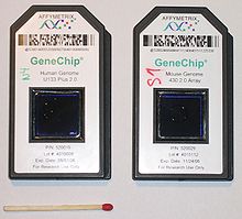
The techniques commonly used for simultaneously measuring the concentration of a large number of different types of mRNA include Microarray, high throughput sequencing said RNA RNA-Seq.
Scientific History
Clues to the existence of a post-mortem transcriptome existed at least since the beginning of the 21st century,[citation needed] but the word thanatotranscriptome seems to have been first used in the scientific literature by Javan et al. in 2015.[2]
In 2016, researchers at the University of Washington confirmed that up to 2 days (48 hours) after the death of mice or zebrafish, many genes still functioned.[1][3] Changes in the quantities of mRNA in the bodies of the dead animals prove that hundreds of genes with very different functions awoke just after death. The researchers detected 548 genes that awoke after death in zebrafish and 515 in laboratory mice. Among these were genes involved in development of the organism, including genes that are normally activated only in utero or in ovo (in the egg) during fetal development.
Applications
This information could possibly in the future lead to:
- Construct a definition both more accurate and nuanced phenomenon of "death".
- More precise time of death by the forensic (or a biologist or veterinarian in an EcoHealth investigation who needs information on hours or causes of poisoning, without the case of a zoonosis for example.
We are far, but if we come to better understand the steps of this phenomenon in the Human, a coroner could via a "postmortem serology"[5] perhaps in the future, according to the dosage of the mRNA, establish with greater precision the time since death (by hour, even in minutes rather than days, which can be useful for investigations to reconstruct the conditions of death[3]). - Illuminate the phenomenon of cell death of apoptosis or the death of a body, and in particular the phenomenon of ischemia (myocardial including[6]) and its process healing or resilience, for perhaps then to facilitate them.
This gene revival also means remaining in the cells for up to 48 hours after the death of these animals enough energy for activating the cellular machinery.[3] At least part of these genes appear to be genes involved in physiological healing, healing or "auto-resuscitation".[3] - Understand cancer. It was found that among the genes reactivated soon after death, some of the genes involved in the process of cancerous (reactivated with a peak of activity reaches about 24 hours after death[3]) ; detailed understanding of this phenomenon could shed light on the phenomenon of carcinogenesis and maybe bring some new elements to better combat.
- Improving the quality of organ transplants. Indeed, the fact that cancer-related genes are activated after the death in animals is information that leads us to consider the time of organ transplantation to reduce the incidence of cancer in people receiving these transplants.[3] Persons to which was grafted a new liver actually more cancers after treatment than would be statistically normal. This phenomenon was attributed to the diet imposed on them, or immunosuppressive drugs they receive for their body does not reject the transplant.[3] One hypothesis (yet to be verified) is that the cancer genes activated in the liver of the donor may also play a role.[3]
- Whether there is the same in humans, because previous studies have already shown that in people dead by trauma, heart attack or suffocation, various genes including those involved in cardiac muscle contraction and wound healing, were active more than 12 hours after death.[6] Similarly for gene dental pulp.[7] Some authors in 2015 introduced the concept of "thanatotranscriptome apoptotic".[2]
- Test another hypothesis is that after death, a rapid decrease of the "suppressor genes" activity (which normally inhibit the activation of other genes, including those no longer needed after the fetal stage) would allow dormant genes wake up, at least for this short period of time.[3]
See also
References
- ^ a b Pozhitkov, Alex E.; Neme, Rafik; Domazet-Lošo, Tomislav; Leroux, Brian G.; Soni, Shivani; Tautz, Diethard; Noble, Peter A. (2017-01-01). "Tracing the dynamics of gene transcripts after organismal death". Open Biology. 7 (1): 160267. doi:10.1098/rsob.160267. ISSN 2046-2441. PMC 5303275. PMID 28123054.
- ^ a b c Javan, Gulnaz T.; Can, Ismail; Finley, Sheree J.; Soni, Shivani (2015-12-01). "The apoptotic thanatotranscriptome associated with the liver of cadavers". Forensic Science, Medicine, and Pathology. 11 (4): 509–516. doi:10.1007/s12024-015-9704-6. ISSN 1556-2891. PMID 26318598. S2CID 21583165.
- ^ a b c d e f g h i j Williams, Anna (21 June 2016). "Hundreds of genes seen sparking to life two days after death". New Scientist. Retrieved 6 March 2022.
- ^ Javan, Gulnaz T.; Finley, Sheree J.; Abidin, Zain; Mulle, Jennifer G. (2016-02-24). "The Thanatomicrobiome: A Missing Piece of the Microbial Puzzle of Death". Frontiers in Microbiology. 7. doi:10.3389/fmicb.2016.00225. ISSN 1664-302X. PMC 4764706. PMID 26941736.
{{cite journal}}: CS1 maint: PMC format (link) CS1 maint: unflagged free DOI (link) - ^ Moreno, L. I., Tate, C. M., Knott, E. L., McDaniel, J. E., Rogers, S. S., Koons, B. W., ... & Robertson, J. M. (2012). Determination of an effective housekeeping gene for the quantification of mRNA for forensic applications. Journal of Forensic Sciences, 57(4), 1051-1058 (summary).
- ^ a b González-Herrera, L., Valenzuela, A., Marchal, J. A., Lorente, J. A., & Villanueva, E. (2013). Studies on RNA integrity and gene expression in human myocardial tissue, pericardial fluid and blood, and its postmortem stability. Forensic science international, 232(1), 218-228 (summary).
- ^ Poór, V. S., Lukács, D., Nagy, T., Rácz, E., & Sipos, K. (2016). The rate of RNA degradation in human dental pulp reveals post-mortem interval. International Journal of Legal Medicine, 130(3), 615-619 (summary).
