
3D Slicer: Difference between revisions
No edit summary |
m formatting journal PMID cites using AWB (7391) |
||
| Line 22: | Line 22: | ||
3D Slicer is a free open source software (BSD license) that is a flexible, modular platform for image analysis and visualization. 3D Slicer can be easily extended to enable development of both interactive and [[batch processing]] tools for a variety of applications. |
3D Slicer is a free open source software (BSD license) that is a flexible, modular platform for image analysis and visualization. 3D Slicer can be easily extended to enable development of both interactive and [[batch processing]] tools for a variety of applications. |
||
3D Slicer provides [[image registration]], processing of [[ |
3D Slicer provides [[image registration]], processing of [[Diffusion MRI|DTI (diffusion tractography)]], an interface to external devices for image guidance support, and [[Gpu|GPU]]-enabled [[volume rendering]], among other capabilities. 3D Slicer has a modular organization that allows the easy addition of new functionality and provides a number of generic features not available in competing tools. |
||
The interactive visualization capabilities of 3D Slicer include the ability to display arbitrarily oriented image slices, build surface models from image labels, and high performance and high performance volume rendering. 3D Slicer also supports a rich set of annotation features ([[ |
The interactive visualization capabilities of 3D Slicer include the ability to display arbitrarily oriented image slices, build surface models from image labels, and high performance and high performance volume rendering. 3D Slicer also supports a rich set of annotation features ([[Fiduciary marker|fiducials]] and measurement widgets, customized colormaps). |
||
Slicer's capabilities include<ref>Pieper S., Lorensen B., Schroeder W., Kikinis R. The NA-MIC Kit: ITK, VTK, Pipelines, Grids and 3D Slicer as an Open Platform for the Medical Image Computing Community. Proceedings of the 3rd IEEE International Symposium on Biomedical Imaging: From Nano to Macro 2006; 1:698-701.</ref>: |
Slicer's capabilities include<ref>Pieper S., Lorensen B., Schroeder W., Kikinis R. The NA-MIC Kit: ITK, VTK, Pipelines, Grids and 3D Slicer as an Open Platform for the Medical Image Computing Community. Proceedings of the 3rd IEEE International Symposium on Biomedical Imaging: From Nano to Macro 2006; 1:698-701.</ref>: |
||
* Handling [[DICOM|DICOM images]] and reading/writing a variety of other formats |
* Handling [[DICOM|DICOM images]] and reading/writing a variety of other formats |
||
* Interactive visualization of [[Voxel|volumertic Voxel images]], [[ |
* Interactive visualization of [[Voxel|volumertic Voxel images]], [[Polygonal modeling|polygonal meshes]], and [[volume rendering]]s |
||
* Manual editing |
* Manual editing |
||
* Fusion and co-registering of data using rigid and non-rigid algorithms |
* Fusion and co-registering of data using rigid and non-rigid algorithms |
||
* Automatic [[ |
* Automatic [[Segmentation (image processing)|image segmentation]] |
||
* Analysis and visualization of diffusion tensor imaging data |
* Analysis and visualization of diffusion tensor imaging data |
||
* Tracking of devices for image-guided procedures. |
* Tracking of devices for image-guided procedures. |
||
| Line 38: | Line 38: | ||
Slicer is compiled for use on multiple platforms, including [[Microsoft Windows|Windows]], [[Linux]], and [[Mac OS X]]. |
Slicer is compiled for use on multiple platforms, including [[Microsoft Windows|Windows]], [[Linux]], and [[Mac OS X]]. |
||
Slicer is distributed under a [[ |
Slicer is distributed under a [[Bsd license|BSD]] style, free, open source license. The license has no restrictions on use of the software. However, no claims are made on the software being useful for any particular task. It is entirely the responsibility of the user to ensure compliance with local rules and regulations. Slicer has not been approved for clinical use in the US or elsewhere. |
||
== Image gallery == |
== Image gallery == |
||
| Line 56: | Line 56: | ||
==History== |
==History== |
||
Slicer started as a masters [[thesis]] project between the Surgical Planning Laboratory at the Brigham and Women's Hospital and the MIT Artificial Intelligence Laboratory in 1998<ref> |
Slicer started as a masters [[thesis]] project between the Surgical Planning Laboratory at the Brigham and Women's Hospital and the MIT Artificial Intelligence Laboratory in 1998<ref>{{cite journal | pmid = 9766770 }}</ref>. 3D Slicer version 2 has been downloaded several thousand times. In 2007 a completely revamped version 3 of Slicer was released. The next major refactoring of Slicer was initiated in 2009, which aims to transition the [[GUI]] of Slicer from using [http://www.kwwidgets.org/Wiki/KWWidgets KWWidgets] to [[Qt]]. Qt-enabled Slicer version 4 is expected to be released in 2010–2011. |
||
Slicer software has enabled a variety of research [[Academic publishing|publications]], all aimed at improving [[image analysis]]<ref>Pieper S., Lorensen B., Schroeder W., Kikinis R. The NA-MIC Kit: ITK, VTK, Pipelines, Grids and 3D Slicer as an Open Platform for the Medical Image Computing Community. Proceedings of the 3rd IEEE International Symposium on Biomedical Imaging: From Nano to Macro 2006; 1:698–701.</ref>. |
Slicer software has enabled a variety of research [[Academic publishing|publications]], all aimed at improving [[image analysis]]<ref>Pieper S., Lorensen B., Schroeder W., Kikinis R. The NA-MIC Kit: ITK, VTK, Pipelines, Grids and 3D Slicer as an Open Platform for the Medical Image Computing Community. Proceedings of the 3rd IEEE International Symposium on Biomedical Imaging: From Nano to Macro 2006; 1:698–701.</ref>. |
||
| Line 66: | Line 66: | ||
Slicer's platform provides functionalities for [[Segmentation (image processing)|segmentation]], [[Image registration|registration]] and [[Three-dimensional space|three-dimensional]] visualization of [[multimodal distribution|multimodal]] image data, as well as advanced image analysis [[algorithms]] for [[diffusion tensor]] imaging, functional [[magnetic resonance imaging]] and [[image-guided radiation therapy]]. Standard [[image file formats]] are supported, and the application integrates interface capabilities to biomedical research software. |
Slicer's platform provides functionalities for [[Segmentation (image processing)|segmentation]], [[Image registration|registration]] and [[Three-dimensional space|three-dimensional]] visualization of [[multimodal distribution|multimodal]] image data, as well as advanced image analysis [[algorithms]] for [[diffusion tensor]] imaging, functional [[magnetic resonance imaging]] and [[image-guided radiation therapy]]. Standard [[image file formats]] are supported, and the application integrates interface capabilities to biomedical research software. |
||
Slicer has been used in a variety of [[clinical research]]. In image-guided therapy research, Slicer is frequently used to construct and visualize collections of MRI data that are available pre- and intra-operatively to allow for the acquiring of spatial [[Coordinate system|coordinates]] for instrument tracking<ref> |
Slicer has been used in a variety of [[clinical research]]. In image-guided therapy research, Slicer is frequently used to construct and visualize collections of MRI data that are available pre- and intra-operatively to allow for the acquiring of spatial [[Coordinate system|coordinates]] for instrument tracking<ref>{{cite journal | pmid = 18051095 }}</ref>. In fact, Slicer has already played such a pivotal role in image-guided therapy, it can be considered as growing up alongside that field, with over 200 publications referencing Slicer since 1998<ref>For a list of publications citing Slicer usage since 1998, visit: http://www.slicer.org/publications/pages/display/?collectionid=11</ref>. |
||
In addition to producing 3D models from conventional MRI images, Slicer has also been used to present information derived from fMRI (using MRI to assess blood flow in the brain related to [[Nervous system|neural]] or [[spinal cord]] activity)<ref> |
In addition to producing 3D models from conventional MRI images, Slicer has also been used to present information derived from fMRI (using MRI to assess blood flow in the brain related to [[Nervous system|neural]] or [[spinal cord]] activity)<ref>{{cite journal | pmid = 17289403 }}</ref>, DTI (using MRI to measure the restricted diffusion of water in imaged tissue)<ref>{{cite journal | pmid = 17354847 }}</ref>, and [[electrocardiography]]<ref>{{cite journal | pmid = 16512925 }}</ref>. For example, Slicer's DTI package allows the conversion and analysis of DTI images. The results of such analysis can be integrated with the results from analysis of [[Morphology (biology)|morphologic]] MRI, MR [[Angiography|angiograms]] and fMRI. Other uses of Slicer include [[paleontology]]<ref>http://openpaleo.blogspot.com/2009/03/3d-slicer-tutorial-part-vi.html</ref> and neurosurgery planning<ref>http://picasaweb.google.com/107065747472066371420</ref>. |
||
==Developers== |
==Developers== |
||
| Line 83: | Line 83: | ||
*[[CMake]] |
*[[CMake]] |
||
*[[CPack]] |
*[[CPack]] |
||
*[[ |
*[[Python (programming language)|Python]] |
||
*[[Tcl]] |
*[[Tcl]] |
||
*[http://teem.sourceforge.net/nrrd/ nrrd] |
*[http://teem.sourceforge.net/nrrd/ nrrd] |
||
Revision as of 20:04, 14 November 2010
 | |
| Original author(s) | The Slicer Community |
|---|---|
| Stable release | 3.6
/ June, 2010 |
| Operating system | Cross-platform |
| Available in | C++, Tcl, Python, Java |
| Type | Scientific visualization and image computing |
| License | BSD-style |
| Website | www.slicer.org |
3D Slicer (Slicer) is a free, open source software package for image analysis[1] and scientific visualization. Slicer is used in a variety of medical applications, including autism, multiple sclerosis, systemic lupus erythematosus, prostate cancer, schizophrenia, orthopedic biomechanics, COPD, cardiovascular disease and neurosurgery.
About Slicer
3D Slicer is a free open source software (BSD license) that is a flexible, modular platform for image analysis and visualization. 3D Slicer can be easily extended to enable development of both interactive and batch processing tools for a variety of applications.
3D Slicer provides image registration, processing of DTI (diffusion tractography), an interface to external devices for image guidance support, and GPU-enabled volume rendering, among other capabilities. 3D Slicer has a modular organization that allows the easy addition of new functionality and provides a number of generic features not available in competing tools.
The interactive visualization capabilities of 3D Slicer include the ability to display arbitrarily oriented image slices, build surface models from image labels, and high performance and high performance volume rendering. 3D Slicer also supports a rich set of annotation features (fiducials and measurement widgets, customized colormaps).
Slicer's capabilities include[2]:
- Handling DICOM images and reading/writing a variety of other formats
- Interactive visualization of volumertic Voxel images, polygonal meshes, and volume renderings
- Manual editing
- Fusion and co-registering of data using rigid and non-rigid algorithms
- Automatic image segmentation
- Analysis and visualization of diffusion tensor imaging data
- Tracking of devices for image-guided procedures.
Slicer is compiled for use on multiple platforms, including Windows, Linux, and Mac OS X.
Slicer is distributed under a BSD style, free, open source license. The license has no restrictions on use of the software. However, no claims are made on the software being useful for any particular task. It is entirely the responsibility of the user to ensure compliance with local rules and regulations. Slicer has not been approved for clinical use in the US or elsewhere.
Image gallery
-
 Hardware accelerated volume rendering with nVidia drivers, (on Windows and Linux only).
 -
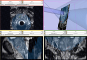 ProstateNav Module for MRI guided robot assisted biopsy of the prostate.
 -
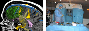 Left: 3D rendering. Right: Open MR system
 -
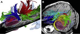
 Visualization of some atlas-based ROIs which correspond to major anatomical fiber tracts. The atlas was provided as part of a download of DTI studio.
 -
 High resolution data acquired on 3-Tesla magnet and post-processed using automated tracking procedure.
 -
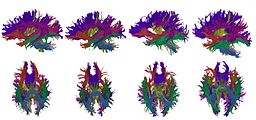 High-dimensional white matter atlas generation and group analysis: result of automatic segmentation of novel subjects.
 -
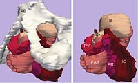
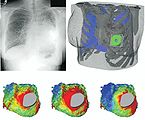 Patient-specific modeling in a patient with congenital heart disease.
 -
 Left: Three-dimensional model of levator ani subdivisions including the pubic bone and pelvic viscera. Right: The same model without the pubic bone.
 -
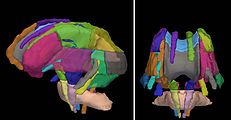
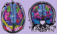 Cortical parcellations derived from SPGR images obtained from a tumor patient.
 -
 Intraoperative colocalization using iMRI images and 3-D Slicer software.


History
Slicer started as a masters thesis project between the Surgical Planning Laboratory at the Brigham and Women's Hospital and the MIT Artificial Intelligence Laboratory in 1998[3]. 3D Slicer version 2 has been downloaded several thousand times. In 2007 a completely revamped version 3 of Slicer was released. The next major refactoring of Slicer was initiated in 2009, which aims to transition the GUI of Slicer from using KWWidgets to Qt. Qt-enabled Slicer version 4 is expected to be released in 2010–2011.
Slicer software has enabled a variety of research publications, all aimed at improving image analysis[4].
This significant software project has been enabled by the participation of several large-scale NIH funded efforts, including the NA-MIC, NAC, BIRN, CIMIT, Harvard Catalyst and NCIGT communities. The funding support comes from several federal funding sources, including NCRR, NIBIB, NIH Roadmap, NCI, NSF and the DOD.
Users
Slicer's platform provides functionalities for segmentation, registration and three-dimensional visualization of multimodal image data, as well as advanced image analysis algorithms for diffusion tensor imaging, functional magnetic resonance imaging and image-guided radiation therapy. Standard image file formats are supported, and the application integrates interface capabilities to biomedical research software.
Slicer has been used in a variety of clinical research. In image-guided therapy research, Slicer is frequently used to construct and visualize collections of MRI data that are available pre- and intra-operatively to allow for the acquiring of spatial coordinates for instrument tracking[5]. In fact, Slicer has already played such a pivotal role in image-guided therapy, it can be considered as growing up alongside that field, with over 200 publications referencing Slicer since 1998[6].
In addition to producing 3D models from conventional MRI images, Slicer has also been used to present information derived from fMRI (using MRI to assess blood flow in the brain related to neural or spinal cord activity)[7], DTI (using MRI to measure the restricted diffusion of water in imaged tissue)[8], and electrocardiography[9]. For example, Slicer's DTI package allows the conversion and analysis of DTI images. The results of such analysis can be integrated with the results from analysis of morphologic MRI, MR angiograms and fMRI. Other uses of Slicer include paleontology[10] and neurosurgery planning[11].
Developers
Slicer is based on VTK, a graphical library that provides a high-level interface to OpenGL and a pipeline mechanism to connect graphical filters. The library is implemented in C++ but provides a Tcl wrapper to instantiate and execute its methods. Tcl/Tk comprises the rest of 3D Slicer user interface and event handling.
Slicer software supports automatic testing and employs an extreme programming approach with nightly builds natively on multiple platforms. Recent accomplishments include added capability for plugging-in external modules using an XML-based command line interface. The Slicer effort also incorporates a light-weight user-centered design effort to promote overall software usability.
Slicer.org offers resources for those who would like to improve or modify Slicer designs and applications. Various tutorials and instructions, along with access to a community of other developers can be found there.
Components of Slicer
External links
References
- ^ Pieper S., Halle M., Kikinis R. 3D SLICER. Proceedings of the 1st IEEE International Symposium on Biomedical Imaging: From Nano to Macro 2004; 1:632–635.
- ^ Pieper S., Lorensen B., Schroeder W., Kikinis R. The NA-MIC Kit: ITK, VTK, Pipelines, Grids and 3D Slicer as an Open Platform for the Medical Image Computing Community. Proceedings of the 3rd IEEE International Symposium on Biomedical Imaging: From Nano to Macro 2006; 1:698-701.
- ^ . PMID 9766770.
{{cite journal}}: Cite journal requires|journal=(help); Missing or empty|title=(help) - ^ Pieper S., Lorensen B., Schroeder W., Kikinis R. The NA-MIC Kit: ITK, VTK, Pipelines, Grids and 3D Slicer as an Open Platform for the Medical Image Computing Community. Proceedings of the 3rd IEEE International Symposium on Biomedical Imaging: From Nano to Macro 2006; 1:698–701.
- ^ . PMID 18051095.
{{cite journal}}: Cite journal requires|journal=(help); Missing or empty|title=(help) - ^ For a list of publications citing Slicer usage since 1998, visit: http://www.slicer.org/publications/pages/display/?collectionid=11
- ^ . PMID 17289403.
{{cite journal}}: Cite journal requires|journal=(help); Missing or empty|title=(help) - ^ . PMID 17354847.
{{cite journal}}: Cite journal requires|journal=(help); Missing or empty|title=(help) - ^ . PMID 16512925.
{{cite journal}}: Cite journal requires|journal=(help); Missing or empty|title=(help) - ^ http://openpaleo.blogspot.com/2009/03/3d-slicer-tutorial-part-vi.html
- ^ http://picasaweb.google.com/107065747472066371420