Extensor indicis muscle
| Extensor indicis proprius | |
|---|---|
 | |
 Posterior surface of the forearm. Deep muscles. Extensor indicis muscle is labeled in purple. | |
| Details | |
| Origin | ulna, interosseous membrane |
| Insertion | index finger (extensor hood) |
| Artery | posterior interosseous artery |
| Nerve | posterior interosseous nerve |
| Actions | extends index finger, wrist |
| Identifiers | |
| Latin | musculus extensor indicis |
| TA98 | A04.6.02.052 |
| TA2 | 2515 |
| FMA | 38524 |
| Anatomical terms of muscle | |
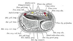
In human anatomy, the extensor indicis [proprius] is a narrow, elongated skeletal muscle in the deep layer of the dorsal forearm, placed medial to, and parallel with, the extensor pollicis longus. Its tendon goes to the index finger, which it extends.
Origin and insertion
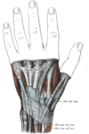
It arises from the distal third of the dorsal part of the body of ulna and from the interosseous membrane. It runs through the fourth tendon compartment together with the extensor digitorum, from where it projects into the dorsal aponeurosis of the index finger. [1]
Opposite the head of the second metacarpal bone, it joins the ulnar side of the tendon of the extensor digitorum which belongs to the index finger.
Like the extensor digiti minimi (i.e. the extensor of the little finger), the tendon of the extensor indicis always runs on the ulnar side of the tendon of the common extensor digitorum. Both these extensors lack the oblique bands (connexus intertendinei) interlinking the tendons of the extensor digitorum on the dorsal side of the hand. [2]
Action
The extensor indicis extends the index finger, and by its continued action assists in extending (dorsiflexion) the wrist and the midcarpal joints[1].
Because the index finger and little finger have separate extensors, these fingers can be moved more independently than the other fingers. [2]
Additional images
Notes
- ^ a b Platzer 2004, p. 168
- ^ a b Ross & Lamperti 2006, p. 300
References
- Platzer, Werner (2004). Color Atlas of Human Anatomy, Vol. 1: Locomotor System (5th ed.). Thieme. ISBN 3-13-533305-1.
{{cite book}}: Invalid|ref=harv(help) - Ross, Lawrence M. (ed.); Lamperti, Edward D. (ed.) (2006). Thieme Atlas of Anatomy: General Anatomy and Musculoskeletal System. Thieme. ISBN 1-58890-419-9.
{{cite book}}:|first1=has generic name (help); Invalid|ref=harv(help)
![]() This article incorporates text in the public domain from page 456 of the 20th edition of Gray's Anatomy (1918)
This article incorporates text in the public domain from page 456 of the 20th edition of Gray's Anatomy (1918)
External links
- Template:MuscleLoyola
- Anatomy photo:09:05-0106 at the SUNY Downstate Medical Center - "Extensor Region of Forearm and Dorsum of Hand: Deep Muscles of Extensor Region"
- lesson5musofpostforearm at The Anatomy Lesson by Wesley Norman (Georgetown University)
- Extensor indicis at the Duke University Health System's Orthopedics program
- Ritter, Merrill A.; Inglis, Allen E. (1969). "The Extensor Indicis Proprius Syndrome" (PDF). J Bone Joint Surg Am. (51): 1645–1648.


