
Sympathetic trunk
This article needs additional citations for verification. (March 2008) |
| Sympathetic trunk | |
|---|---|
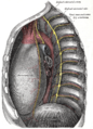
 Abdominal portion of the sympathetic trunk, with the celiac plexus and hypogastric plexus. (Sympathetic trunk labeled at center left.) | |
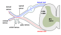
 Scheme showing pathways (white/grey rami are spatially reversed, possibly for clarity?) of a typical spinal nerve. 1. Somatic efferent. 2. Somatic afferent. 3,4,5. Sympathetic efferent. 6,7. Parasympathetic afferent. | |
| Details | |
| Identifiers | |
| Latin | truncus sympathicus |
| TA98 | A14.3.01.002 |
| TA2 | 6602 |
| FMA | 6258 |
| Anatomical terms of neuroanatomy | |
The sympathetic trunks (sympathetic chain, gangliated cord) are a paired bundle of nerve fibers that run from the base of the skull to the coccyx.
Structure
The sympathetic trunk travels in a downward direction from the skull, just lateral to the vertebral bodies. It interacts with the spinal nerves or their ventral rami by way of rami communicantes.
The superior end of it is continued upward through the carotid canal into the skull, and forms a plexus on the internal carotid artery; the inferior part travels in front of the coccyx, where it converges with the other trunk at a structure known as the ganglion impar.
Along the length of the sympathetic trunk are ganglia known as paravertebral ganglia.
Function
The sympathetic trunk is a fundamental part of the sympathetic division of the autonomic nervous system. It allows nerve fibers to travel to spinal nerves that are superior and inferior to the one in which they originated. Also, a number of nerves, such as most of the splanchnic nerves, arise directly from the trunks.
Additional images
-
 The formation of the spinal nerve from the dorsal and ventral roots.
 -
 Transverse section of thorax, showing relations of pulmonary artery.
 -
 The thoracic aorta, viewed from the left side.
 -
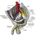
 Dissection of side wall of pelvis showing sacral and pudendal plexuses.
 -
 Sacral plexus of the right side.
 -
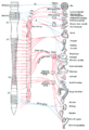 Diagram of efferent sympathetic nervous system.
 -
 Sympathetic connections of the ciliary and superior cervical ganglia.
 -
 Sacral sympathetic
 -
 Sacral sympathetic trunk
 -
 Sacral sympathetic trunk
 -
 Sympathetic cervical trunk


See also
External links
- Anatomy figure: 21:04-04 at Human Anatomy Online, SUNY Downstate Medical Center - "The position of the right and left vagus nerves, and sympathetic trunks in the mediastinum."
- Anatomy photo:43:15-0102 at the SUNY Downstate Medical Center - "The Female Pelvis: The Posterolateral Pelvic Wall"
- . GPnotebook https://www.gpnotebook.co.uk/simplepage.cfm?ID=-1764753348.
{{cite web}}: Missing or empty|title=(help) - Template:EMedicineDictionary
- Atlas image: n3a6p1 at the University of Michigan Health System - "Autonomic Connections of the Spinal Cord"
- Diagram at umm.edu
![]() This article incorporates text in the public domain from page 976 of the 20th edition of Gray's Anatomy (1918)
This article incorporates text in the public domain from page 976 of the 20th edition of Gray's Anatomy (1918)
Anatomy of the autonomic nervous system | |||||
|---|---|---|---|---|---|
| Head |
| ||||
| Neck |
| ||||
| Thorax |
| ||||
| Abdomen |
| ||||
| Pelvis |
| ||||